- Filter By:
-
-
Stock photos and images of central nervous system

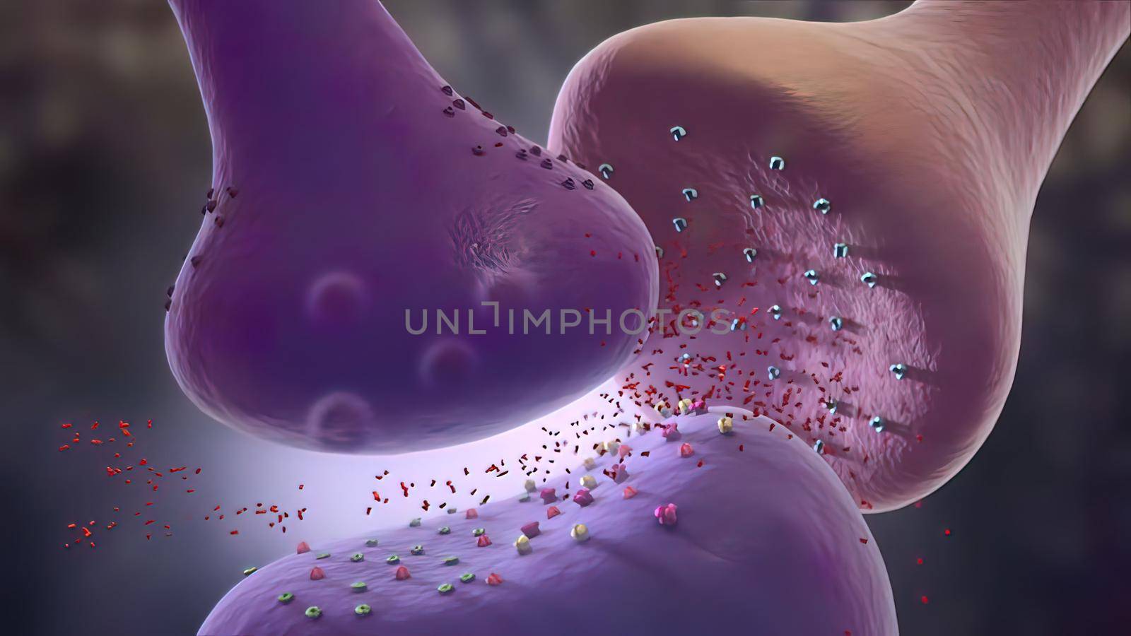
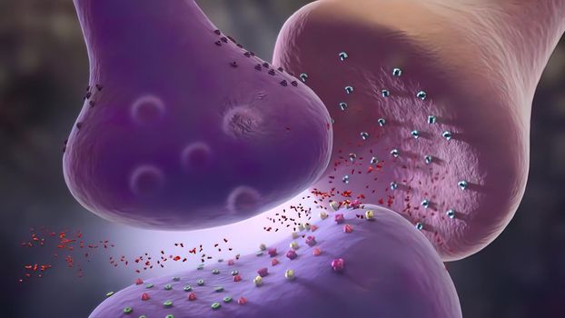
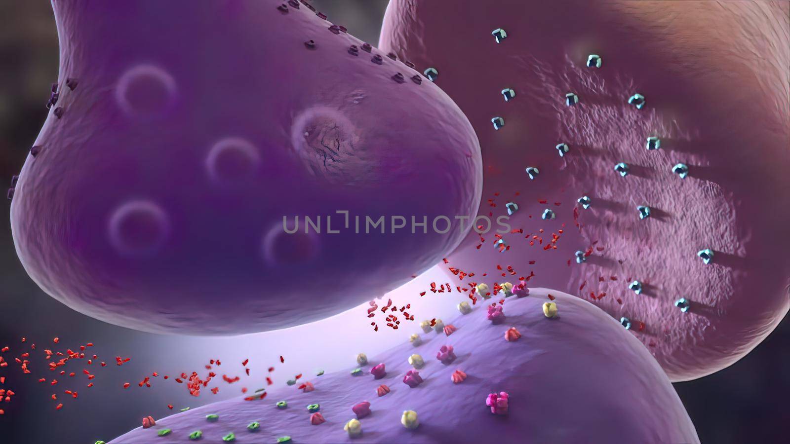
Synaptic transmission, human nervous system
Stock PhotoUsername
creativepicResolution
3840x2160pxSynaptic transmission, human nervous system
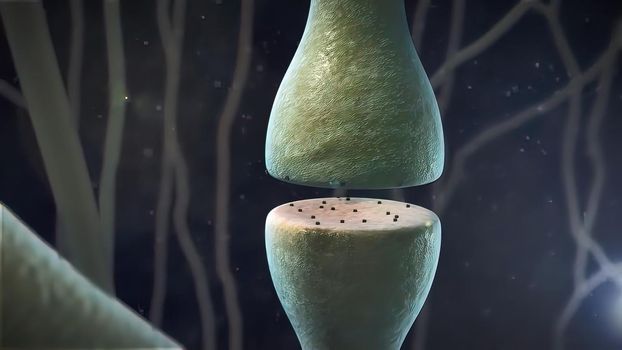
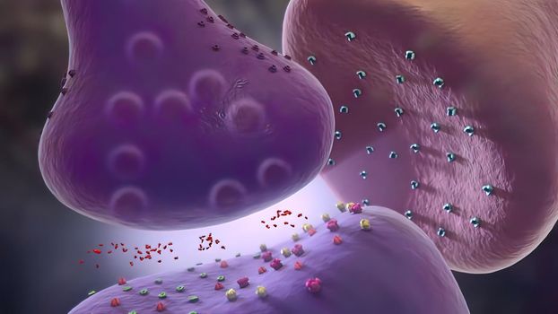
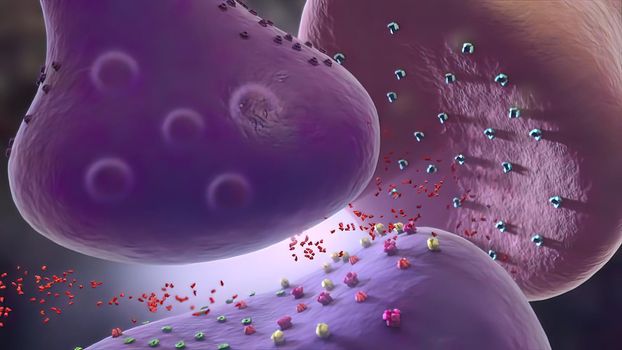
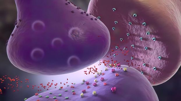
Synaptic transmission, human nervous system
Stock PhotoUsername
creativepicResolution
3840x2160pxSynaptic transmission, human nervous system
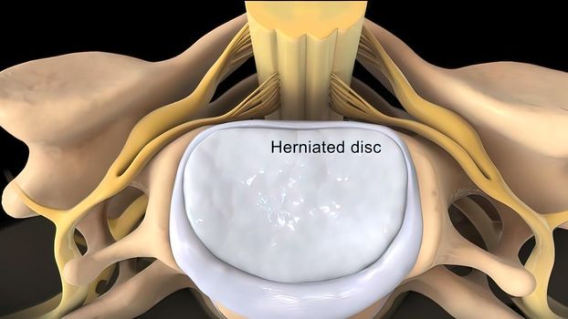
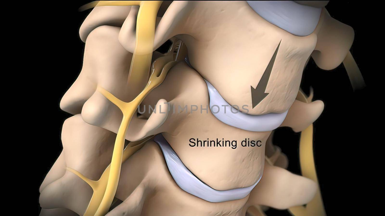
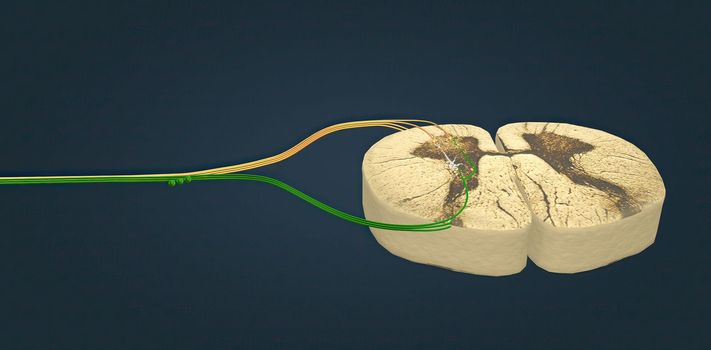
Spinal cord, disc and nervous system
Stock PhotoUsername
creativepicResolution
3840x2160pxSpinal cord, disc and nervous system
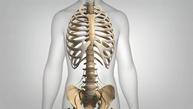
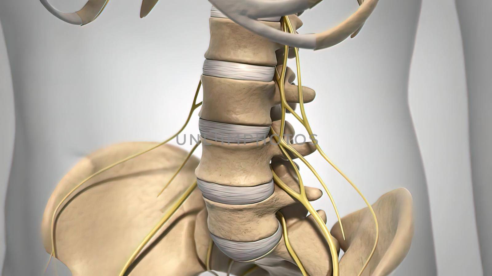
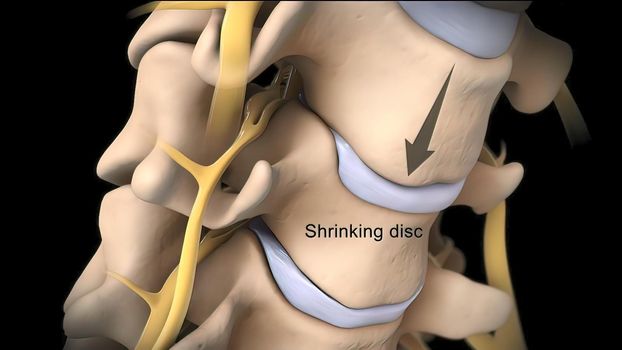
Spinal cord, disc and nervous system
Stock PhotoUsername
creativepicResolution
3840x2160pxSpinal cord, disc and nervous system
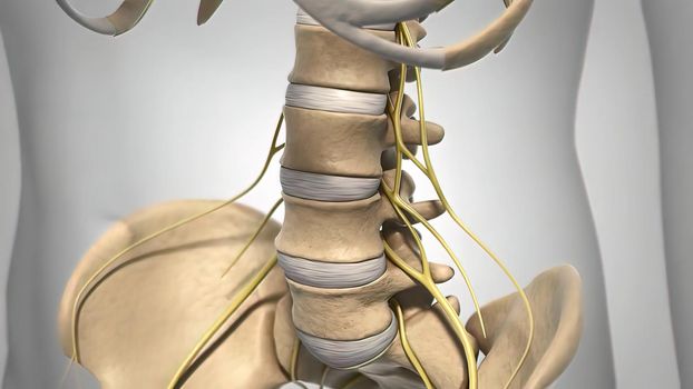

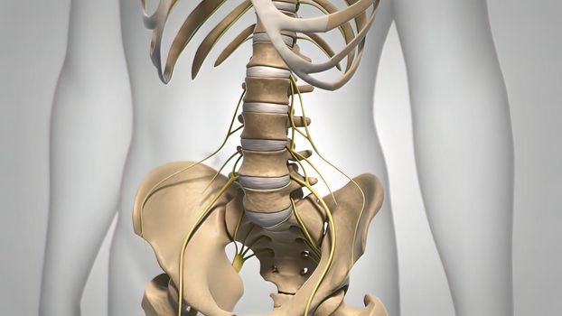
Spinal cord, disc and nervous system
Stock PhotoUsername
creativepicResolution
3840x2160pxSpinal cord, disc and nervous system
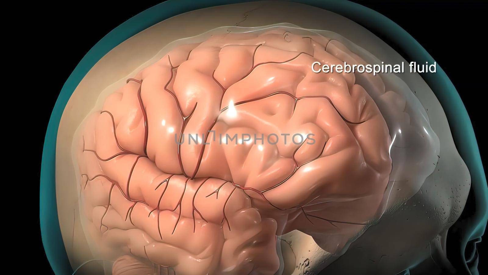
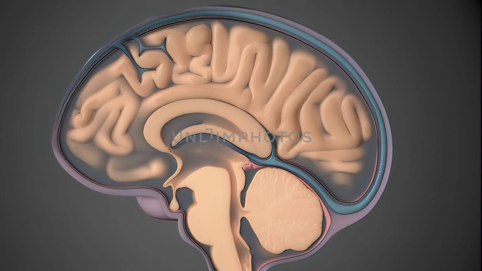
Cerebrospinal fluid is a clear, plasma-like fluid that bathes the central nervous system
Stock PhotoUsername
creativepicResolution
3840x2160pxCerebrospinal fluid is a clear, plasma-like fluid that bathes the central nervous system


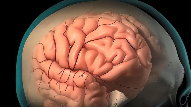
Cerebrospinal fluid is a clear, plasma-like fluid that bathes the central nervous system
Stock PhotoUsername
creativepicResolution
3840x2160pxCerebrospinal fluid is a clear, plasma-like fluid that bathes the central nervous system

Cerebrospinal fluid is a clear, plasma-like fluid that bathes the central nervous system
Stock PhotoUsername
creativepicResolution
3840x2160pxCerebrospinal fluid is a clear, plasma-like fluid that bathes the central nervous system
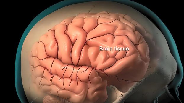

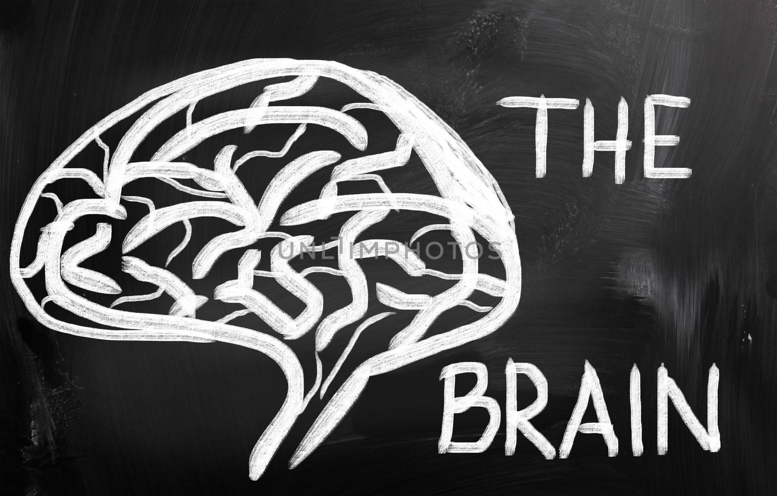
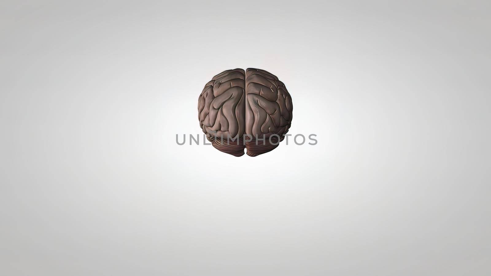
The Structure And Function Of The Human Brain
Stock PhotoUsername
creativepicResolution
3840x2160pxThe Structure And Function Of The Human Brain
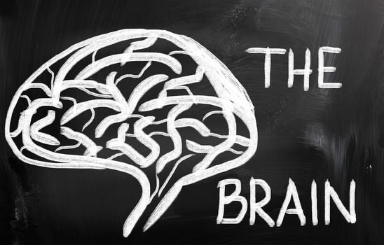
The Structure And Function Of The Human Brain
Stock PhotoUsername
creativepicResolution
3840x2160pxThe Structure And Function Of The Human Brain


The Structure And Function Of The Human Brain
Stock PhotoUsername
creativepicResolution
3840x2160pxThe Structure And Function Of The Human Brain
The human nervous system is destroyed. 3D Medical .
Stock PhotoUsername
creativepicResolution
7680x4320pxThe human nervous system is destroyed. 3D Medical .


The human nervous system is destroyed. 3D Medical .
Stock PhotoUsername
creativepicResolution
7680x4320pxThe human nervous system is destroyed. 3D Medical .

The human nervous system is destroyed. 3D Medical .
Stock PhotoUsername
creativepicResolution
7680x4320pxThe human nervous system is destroyed. 3D Medical .

The human nervous system is destroyed. 3D Medical .
Stock PhotoUsername
creativepicResolution
7680x4320pxThe human nervous system is destroyed. 3D Medical .
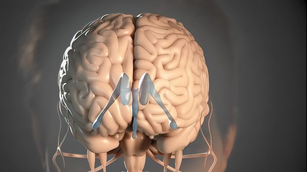
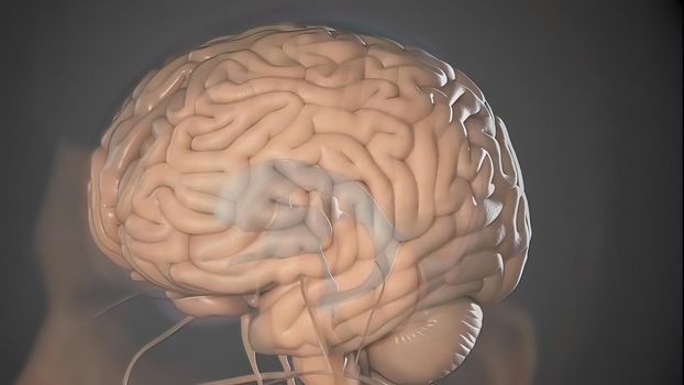
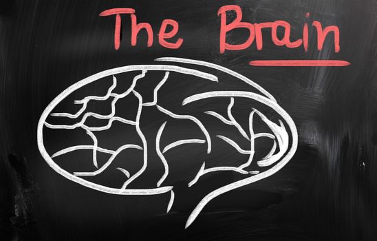
Human Brain
Stock PhotoUsername
KrasimiraNevenovaResolution
4096x2616pxHuman Brain

Human Brain
Stock PhotoUsername
KrasimiraNevenovaResolution
4096x2616pxHuman Brain

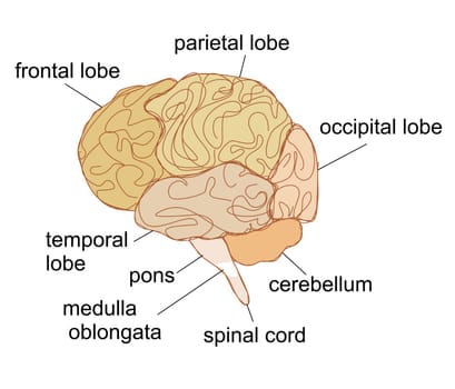
Brain sections vector illustration
Stock PhotoUsername
JackyBrownResolution
5500x4713pxBrain sections vector illustration
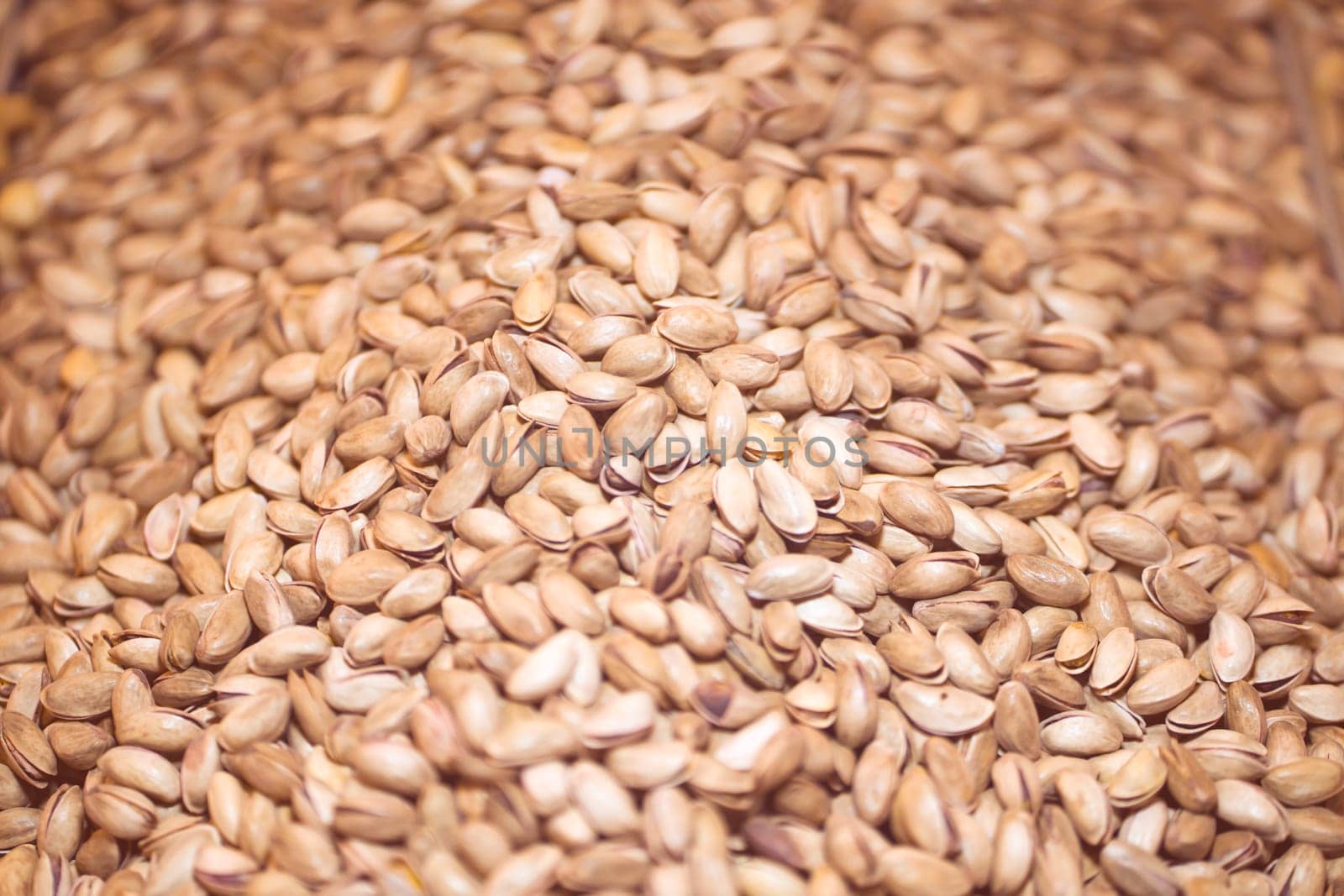
Lots of pistachios, background with tasty nuts.
Stock PhotoUsername
africapinkResolution
6000x4000pxLots of pistachios, background with tasty nuts.


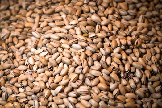
Lots of pistachios, background with tasty nuts.
Stock PhotoUsername
africapinkResolution
5792x3862pxLots of pistachios, background with tasty nuts.

Tramadol pills. Green-yellow capsule pills on blue background. Tramadol is a strong painkiller medicine used to treat moderate to severe pain. Background for tramadol misuse topics. Opioids drug.
Stock PhotoUsername
FahroniResolution
6275x3824pxTramadol pills. Green-yellow capsule pills on blue background. Tramadol is a strong painkiller medicine used to treat moderate to severe pain. Background for tramadol misuse topics. Opioids drug.


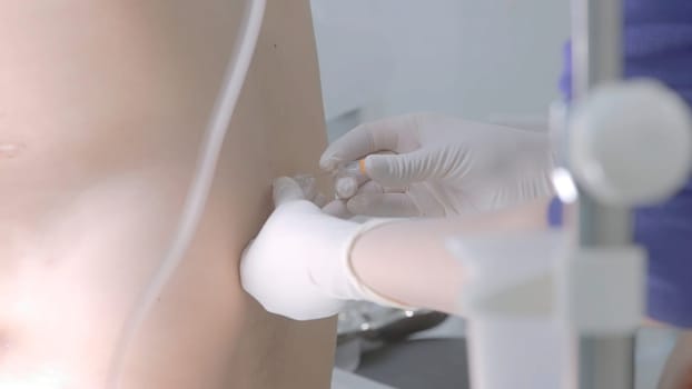
Close up of the process of Lumbar puncture. Action. A medical doctor performing spinal puncture at the hospital.
Stock PhotoUsername
MediawhalestockResolution
7680x4320pxClose up of the process of Lumbar puncture. Action. A medical doctor performing spinal puncture at the hospital.


Close up of the process of Lumbar puncture. Action. A medical doctor performing spinal puncture at the hospital.
Stock PhotoUsername
MediawhalestockResolution
7680x4320pxClose up of the process of Lumbar puncture. Action. A medical doctor performing spinal puncture at the hospital.

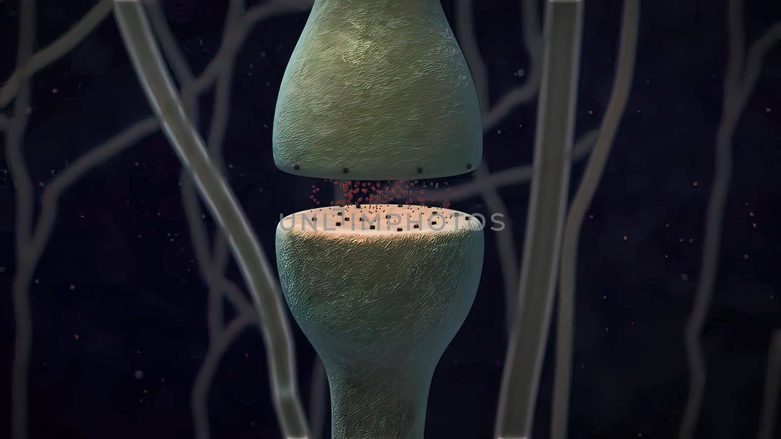
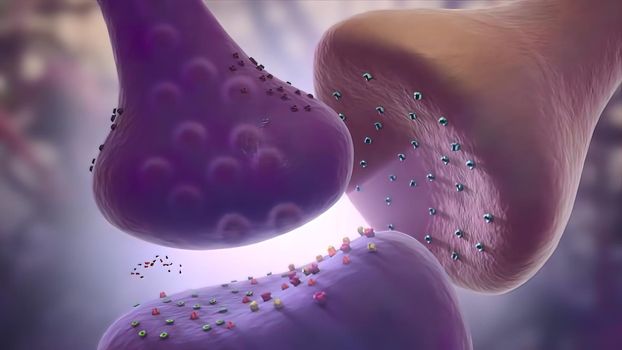
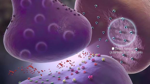
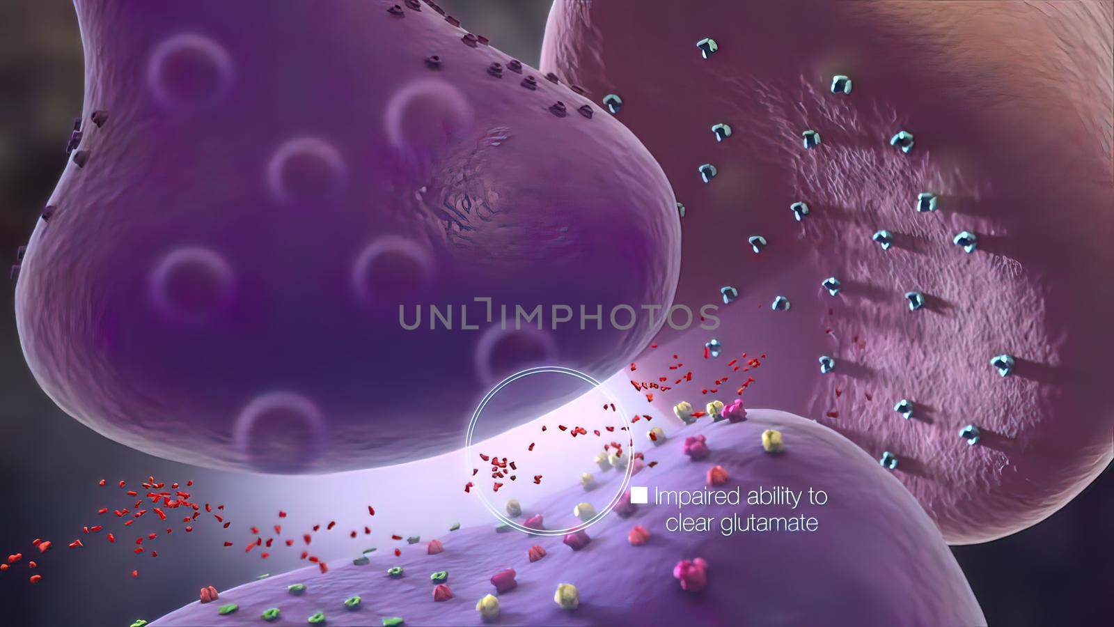
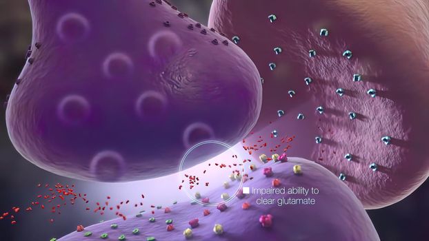
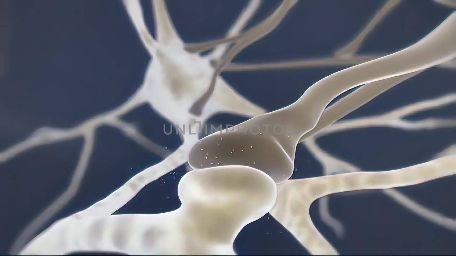
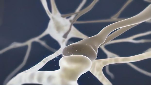
Glial cells of the nervous system release transmitters to release neuronal and synaptic activities.
Stock PhotoUsername
creativepicResolution
3840x2160pxGlial cells of the nervous system release transmitters to release neuronal and synaptic activities.
Glial cells of the nervous system release transmitters to release neuronal and synaptic activities.
Stock PhotoUsername
creativepicResolution
3840x2160pxGlial cells of the nervous system release transmitters to release neuronal and synaptic activities.
Glial cells of the nervous system release transmitters to release neuronal and synaptic activities.
Stock PhotoUsername
creativepicResolution
3840x2160pxGlial cells of the nervous system release transmitters to release neuronal and synaptic activities.

Glial cells of the nervous system release transmitters to release neuronal and synaptic activities.
Stock PhotoUsername
creativepicResolution
3840x2160pxGlial cells of the nervous system release transmitters to release neuronal and synaptic activities.

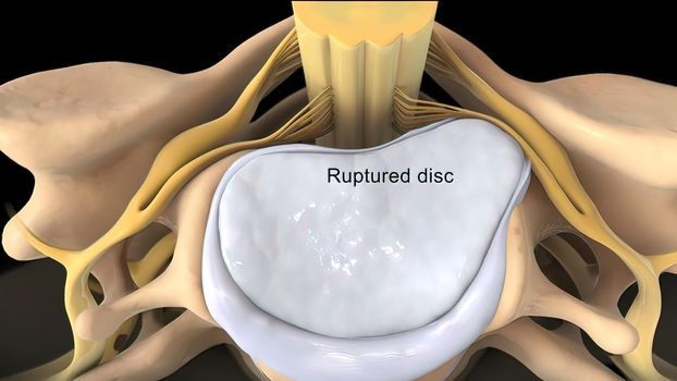
Central disc protrusion top view
Stock PhotoUsername
creativepicResolution
3840x2160pxCentral disc protrusion top view

Central disc protrusion top view
Stock PhotoUsername
creativepicResolution
3840x2160pxCentral disc protrusion top view
Central disc protrusion top view
Stock PhotoUsername
creativepicResolution
3840x2160pxCentral disc protrusion top view
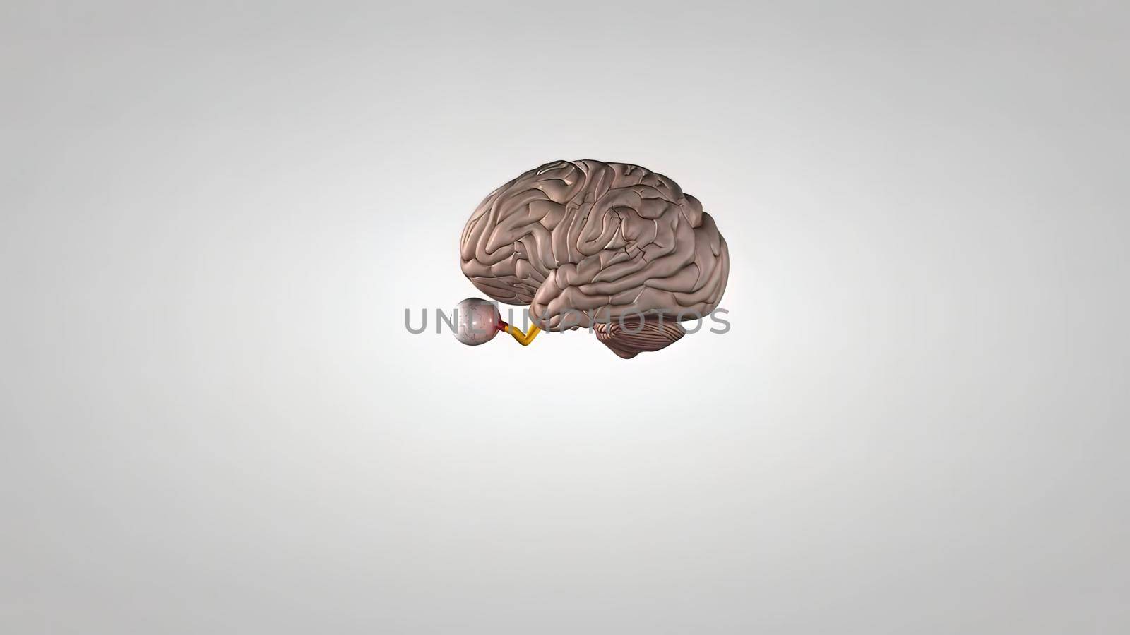
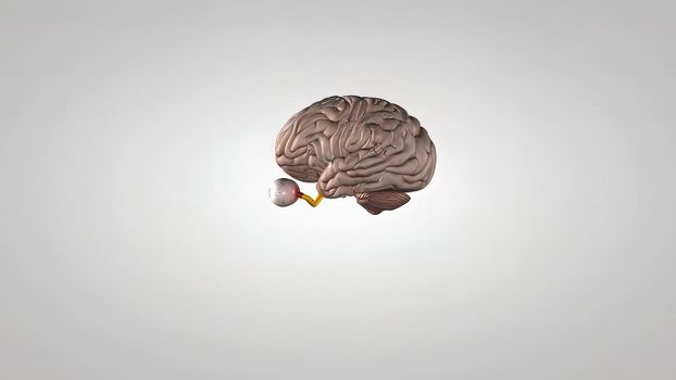
eyes and brain showing the central nervous system, including the brain and eyes
Stock PhotoUsername
creativepicResolution
7680x4320pxeyes and brain showing the central nervous system, including the brain and eyes

eyes and brain showing the central nervous system, including the brain and eyes
Stock PhotoUsername
creativepicResolution
7680x4320pxeyes and brain showing the central nervous system, including the brain and eyes
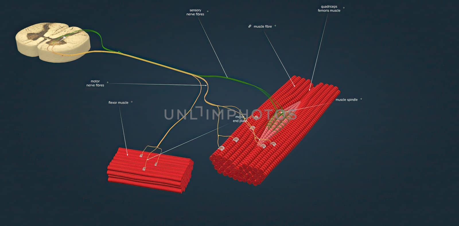
Sensory nerve fibers innervate muscles and tendons, ligaments,
Stock PhotoUsername
creativepicResolution
5482x2700pxSensory nerve fibers innervate muscles and tendons, ligaments,

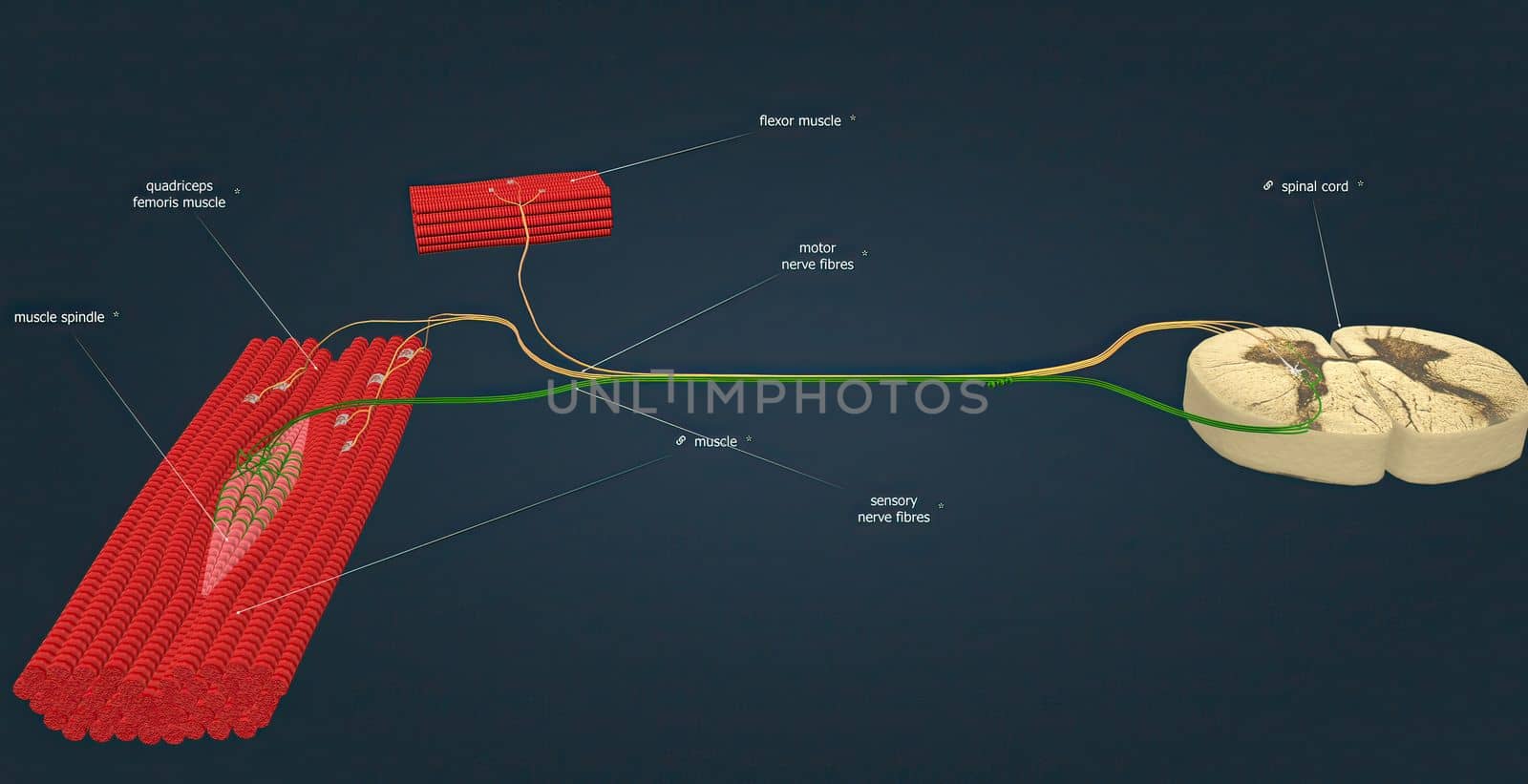
Sensory nerve fibers innervate muscles and tendons,
Stock PhotoUsername
creativepicResolution
5522x2832pxSensory nerve fibers innervate muscles and tendons,


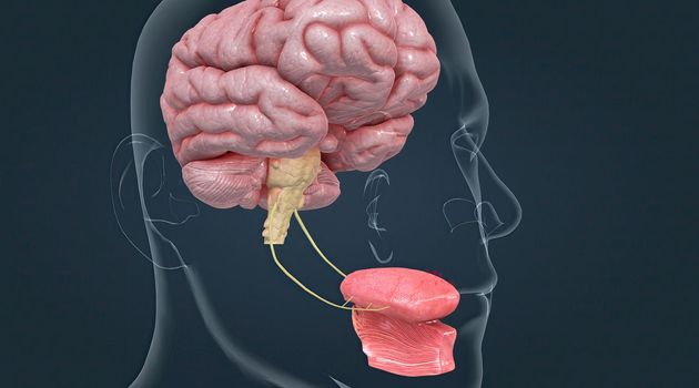
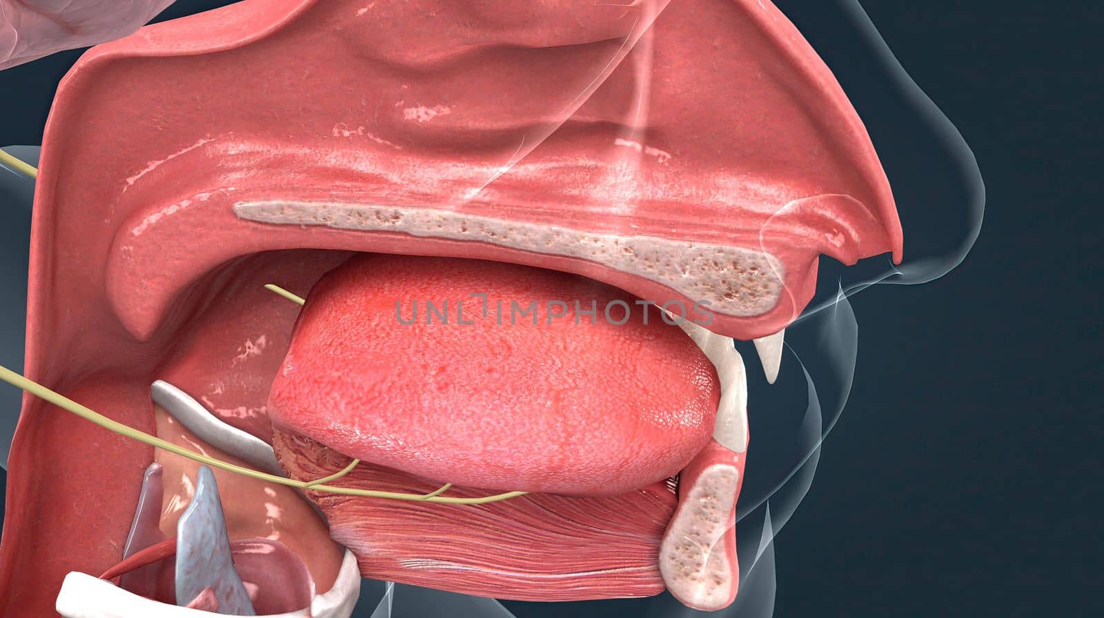
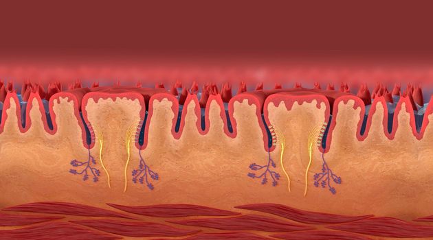
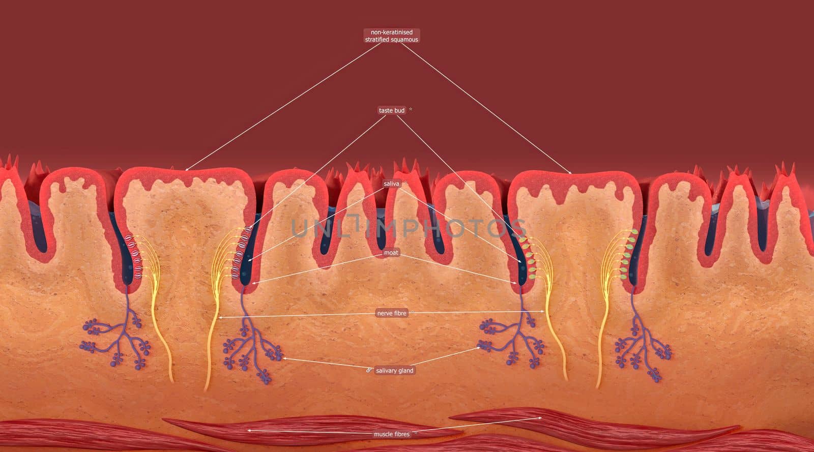
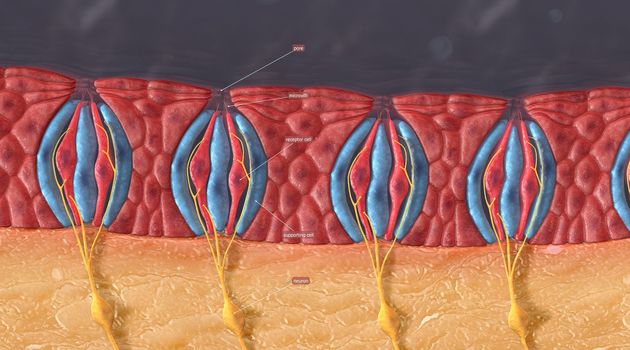
Taste is sensed by chemosensory receptors known as taste buds.
Stock PhotoUsername
creativepicResolution
5404x3002pxTaste is sensed by chemosensory receptors known as taste buds.

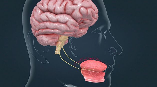
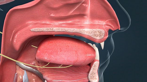
Taste is sensed by chemosensory receptors known as taste buds.
Stock PhotoUsername
creativepicResolution
5404x3002pxTaste is sensed by chemosensory receptors known as taste buds.

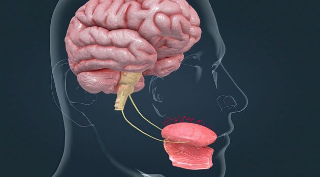
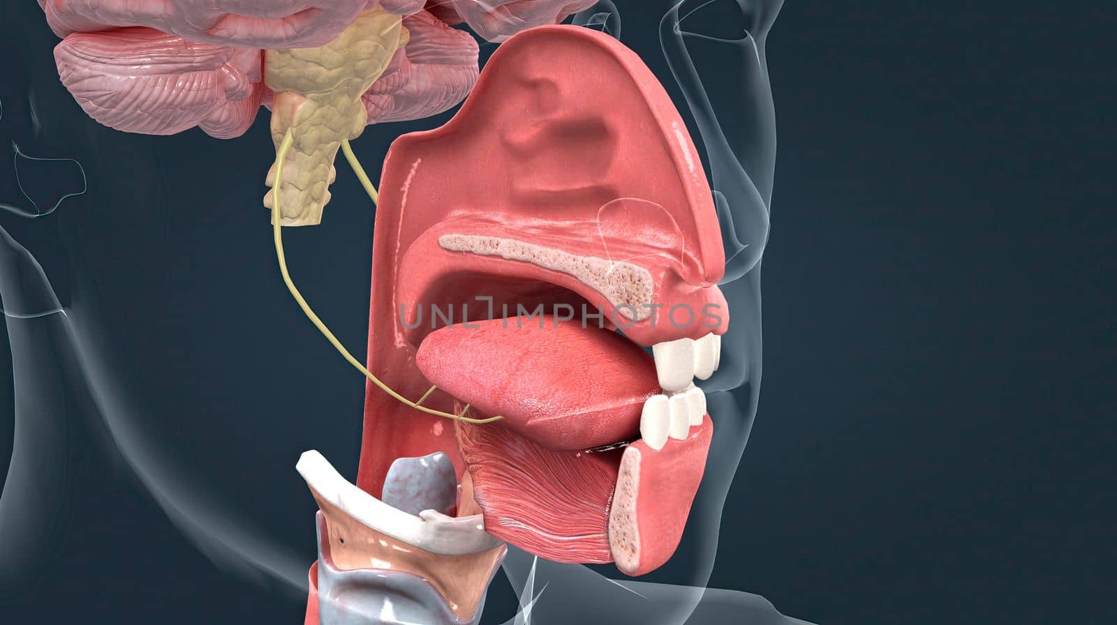
Taste is sensed by chemosensory receptors known as taste buds.
Stock PhotoUsername
creativepicResolution
5404x3002pxTaste is sensed by chemosensory receptors known as taste buds.

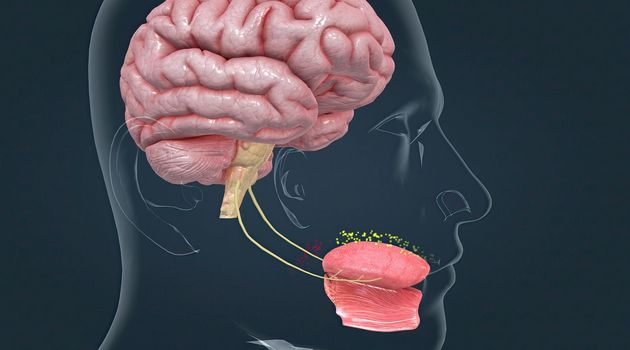
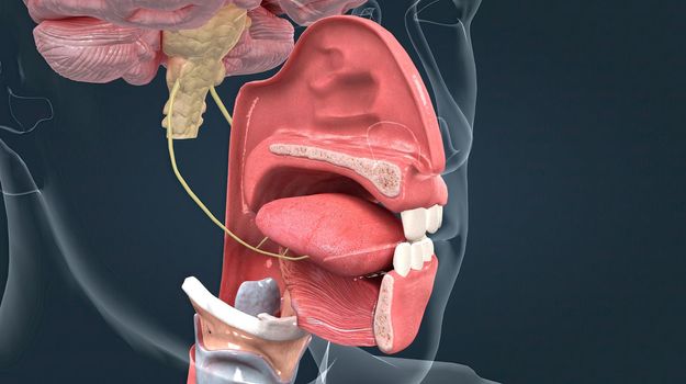
Taste is sensed by chemosensory receptors known as taste buds.
Stock PhotoUsername
creativepicResolution
5404x3002pxTaste is sensed by chemosensory receptors known as taste buds.
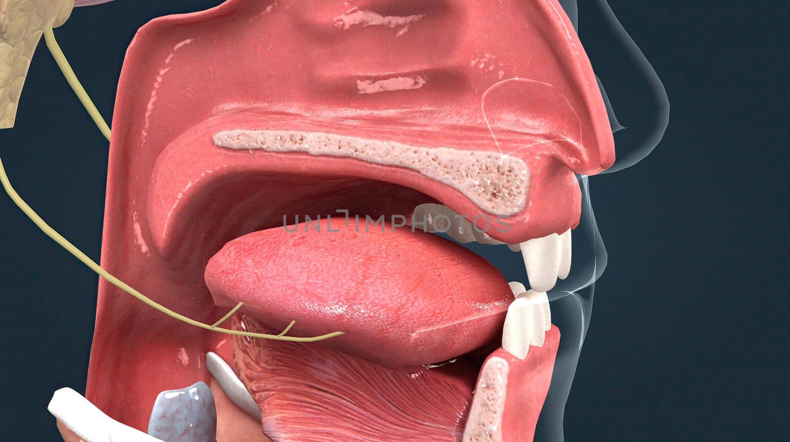
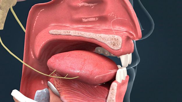
Olfactory organ there are two olfactory bulbs on the bottom side of the brain, one above each nasal cavity.
Stock PhotoUsername
creativepicResolution
5400x3024pxOlfactory organ there are two olfactory bulbs on the bottom side of the brain, one above each nasal cavity.


Olfactory organ there are two olfactory bulbs on the bottom side of the brain, one above each nasal cavity.
Stock PhotoUsername
creativepicResolution
5400x3024pxOlfactory organ there are two olfactory bulbs on the bottom side of the brain, one above each nasal cavity.
Olfactory organ there are two olfactory bulbs on the bottom side of the brain, one above each nasal cavity.
Stock PhotoUsername
creativepicResolution
5400x3024pxOlfactory organ there are two olfactory bulbs on the bottom side of the brain, one above each nasal cavity.
Olfactory organ there are two olfactory bulbs on the bottom side of the brain, one above each nasal cavity.
Stock PhotoUsername
creativepicResolution
5400x3024pxOlfactory organ there are two olfactory bulbs on the bottom side of the brain, one above each nasal cavity.

Lingual papillae are small structures on the upper surface of the tongue that give it its rough texture.
Stock PhotoUsername
creativepicResolution
5404x3002pxLingual papillae are small structures on the upper surface of the tongue that give it its rough texture.
Lingual papillae are small structures on the upper surface of the tongue that give it its rough texture.
Stock PhotoUsername
creativepicResolution
5404x3002pxLingual papillae are small structures on the upper surface of the tongue that give it its rough texture.
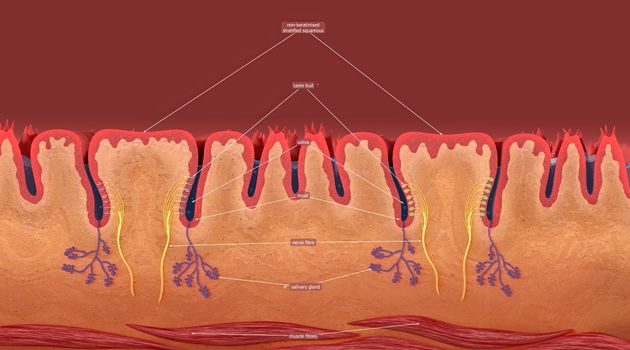
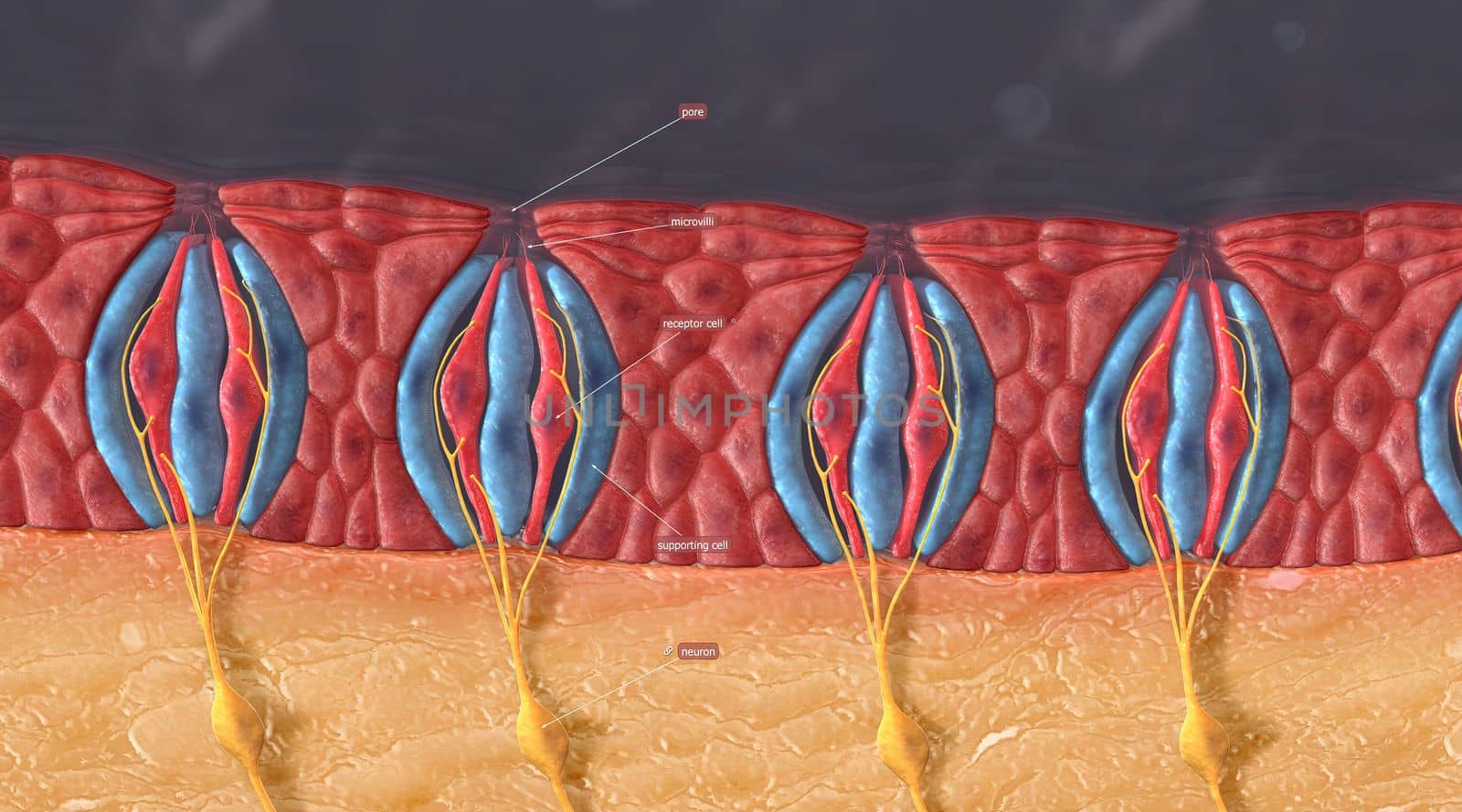
Taste buds contain the taste receptor cells, which are also known as gustatory cells.
Stock PhotoUsername
creativepicResolution
5404x3002pxTaste buds contain the taste receptor cells, which are also known as gustatory cells.
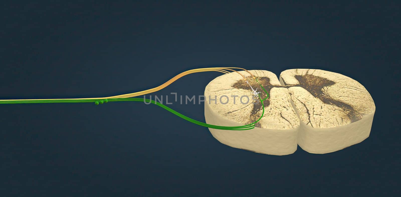
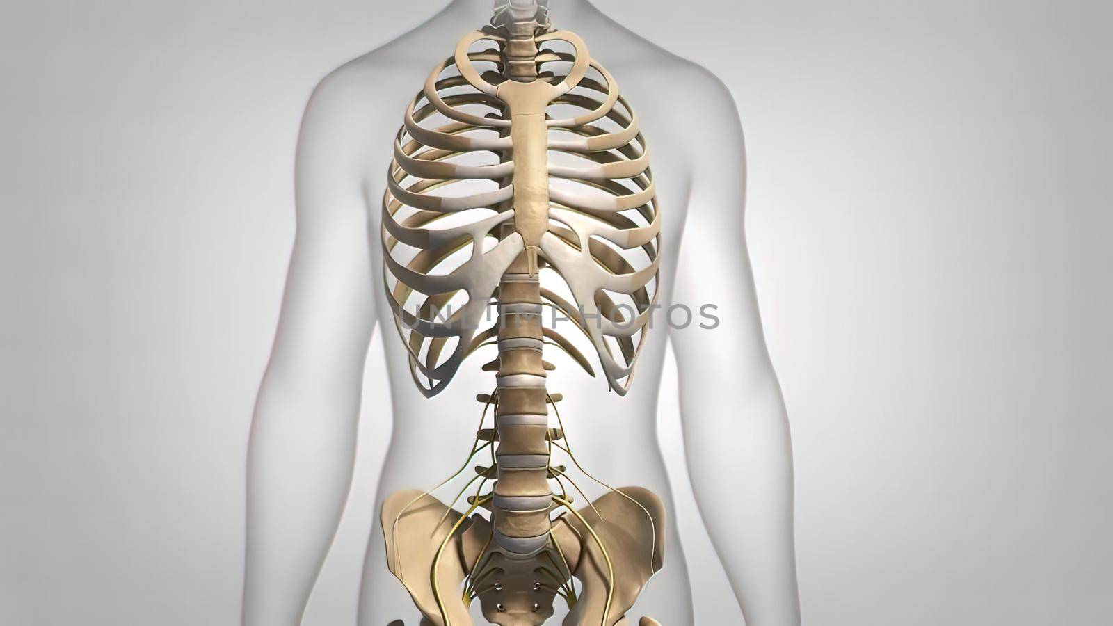
The spinal cord consists of a column of the spine.
Stock PhotoUsername
creativepicResolution
5482x2700pxThe spinal cord consists of a column of the spine.
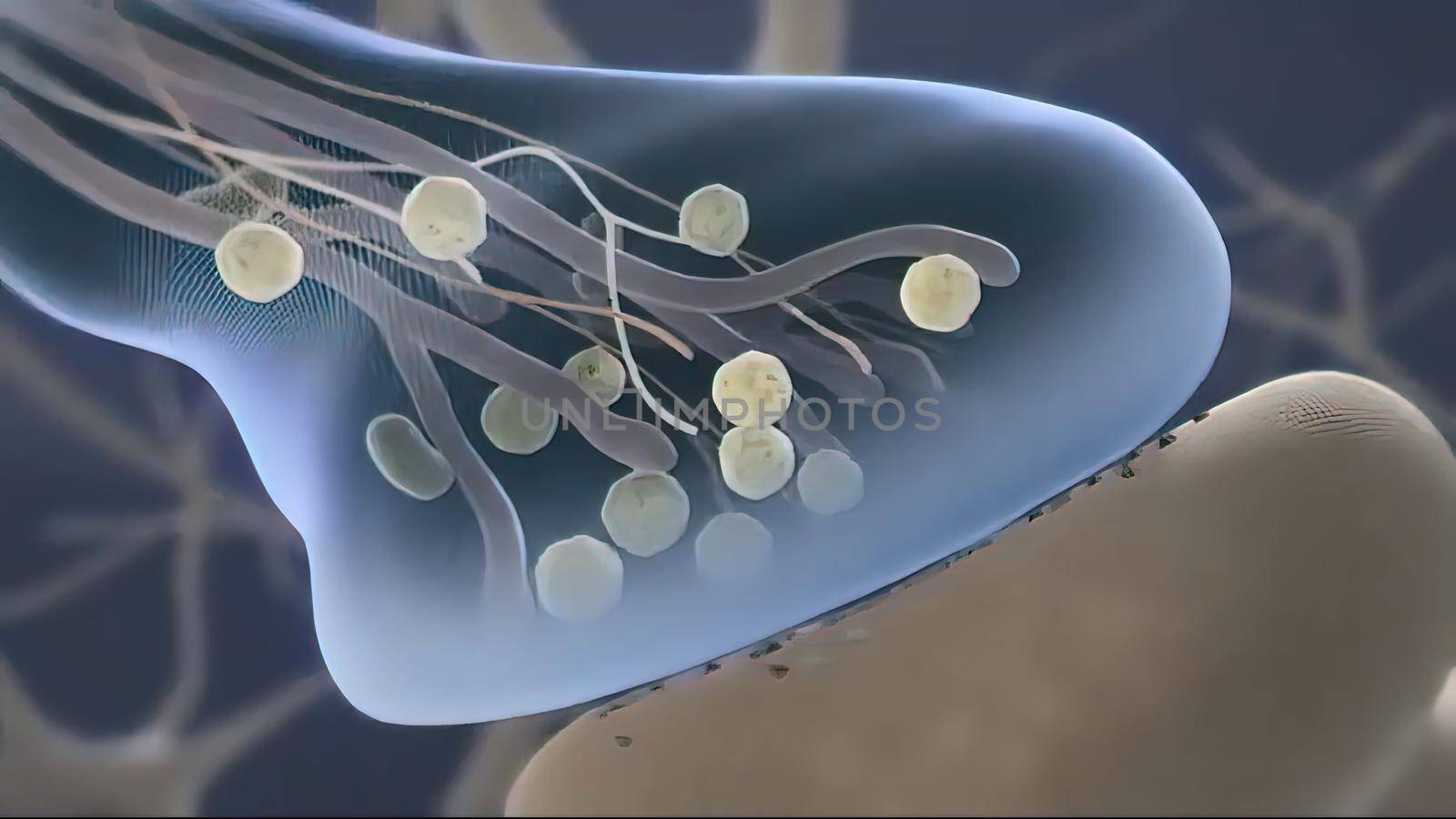
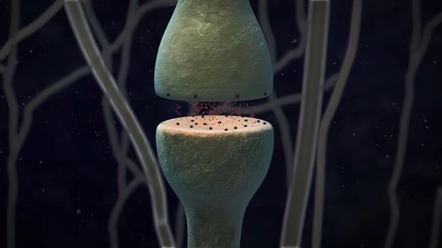
Brain cell synapse showing chemical messengers or neurotransmitters released
Stock PhotoUsername
creativepicResolution
3840x2160pxBrain cell synapse showing chemical messengers or neurotransmitters released

Brain cell synapse showing chemical messengers or neurotransmitters released
Stock PhotoUsername
creativepicResolution
3840x2160pxBrain cell synapse showing chemical messengers or neurotransmitters released
Brain cell synapse showing chemical messengers or neurotransmitters released
Stock PhotoUsername
creativepicResolution
3840x2160pxBrain cell synapse showing chemical messengers or neurotransmitters released
