- Filter By:
-
-
Stock photos and images of username:samunella

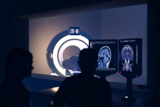
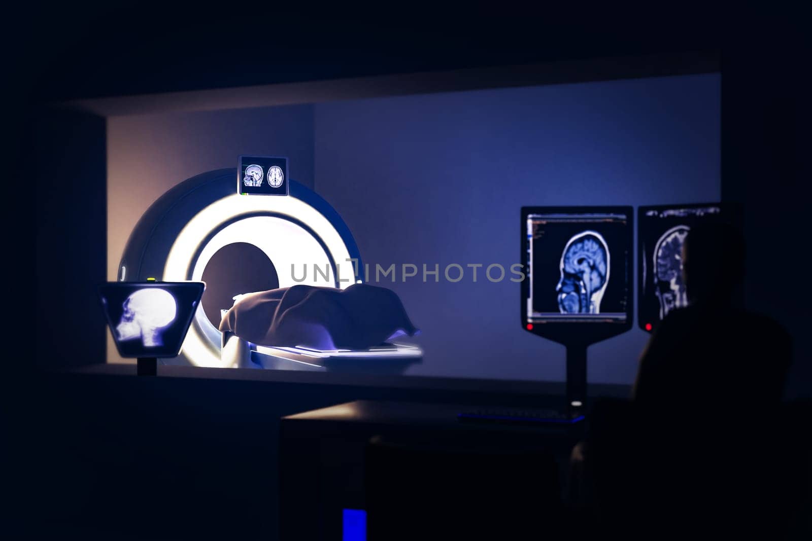
In Control Room Doctor and Radiologist Discuss Diagnosis while Watching Procedure and Monitors Showing Brain Scans Results, In the Background Patient Undergoes MRI or CT Scan Procedure.3D rendering .
Stock PhotoUsername
samunellaResolution
6000x4000pxIn Control Room Doctor and Radiologist Discuss Diagnosis while Watching Procedure and Monitors Showing Brain Scans Results, In the Background Patient Undergoes MRI or CT Scan Procedure.3D rendering .

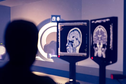
In Control Room Radiologist Diagnosis while Watching Procedure and Monitors Showing Brain Scans Results, In the Background Patient Undergoes MRI or CT Scan Procedure.3D rendering .
Stock PhotoUsername
samunellaResolution
6000x4000pxIn Control Room Radiologist Diagnosis while Watching Procedure and Monitors Showing Brain Scans Results, In the Background Patient Undergoes MRI or CT Scan Procedure.3D rendering .
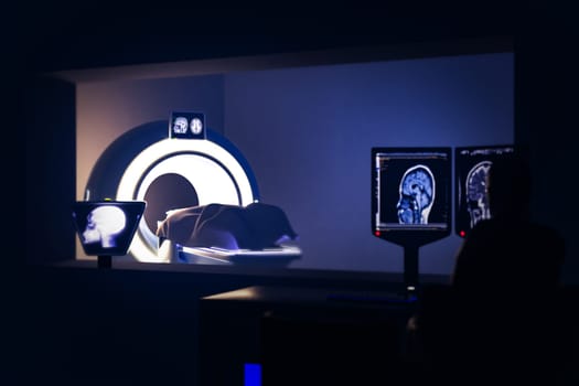
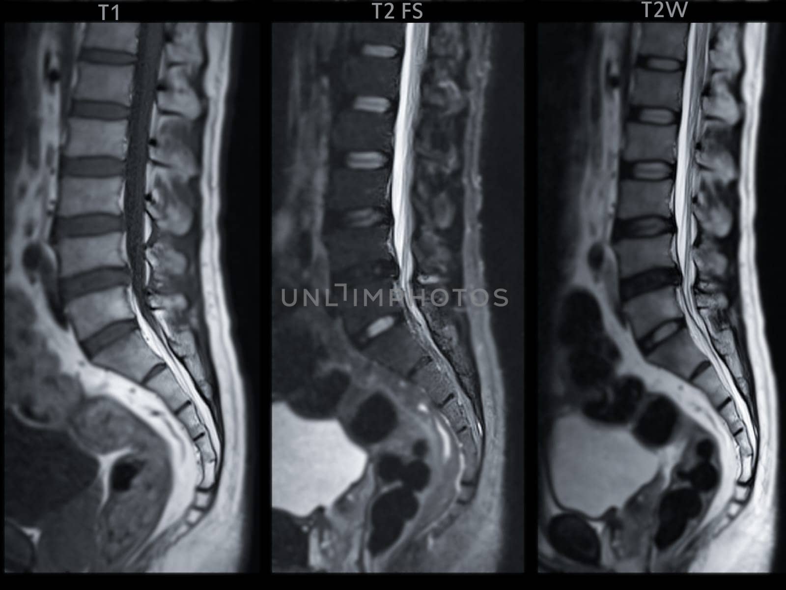
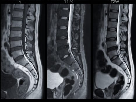
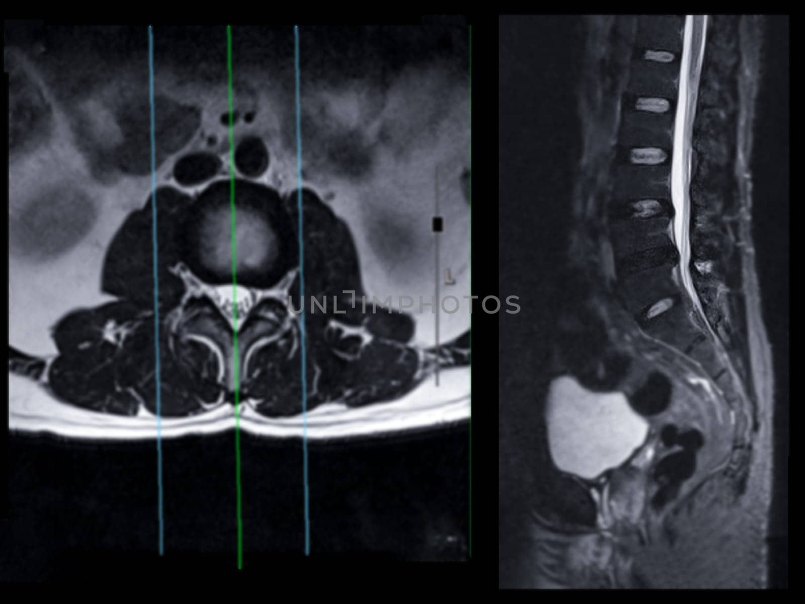
MRI L-S spine or lumbar spine Sagittall T1W ,T2 FS and T2W view for diagnosis spinal cord compression.
Stock PhotoUsername
samunellaResolution
4000x3000pxMRI L-S spine or lumbar spine Sagittall T1W ,T2 FS and T2W view for diagnosis spinal cord compression.
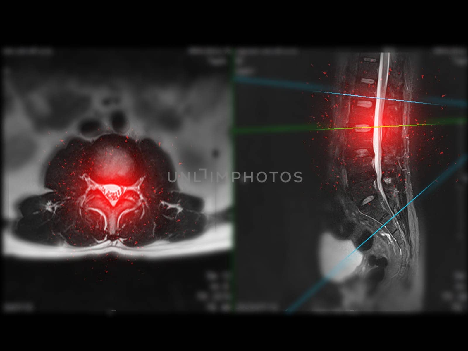
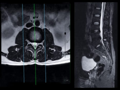
MRI L-S spine or lumbar spine Axial T2W view with sagittal plane for diagnosis spinal cord compression.
Stock PhotoUsername
samunellaResolution
4000x3000pxMRI L-S spine or lumbar spine Axial T2W view with sagittal plane for diagnosis spinal cord compression.
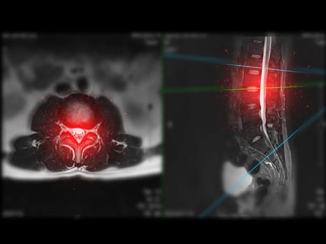

MRI L-S spine or lumbar spine Axial T2W view with sagittal plane for diagnosis spinal cord compression.
Stock PhotoUsername
samunellaResolution
4000x3000pxMRI L-S spine or lumbar spine Axial T2W view with sagittal plane for diagnosis spinal cord compression.
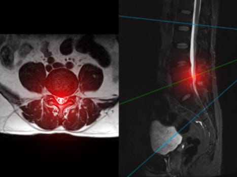
MRI L-S spine or lumbar spine Axial T2W view with sagittal plane for diagnosis spinal cord compression.
Stock PhotoUsername
samunellaResolution
4000x3000pxMRI L-S spine or lumbar spine Axial T2W view with sagittal plane for diagnosis spinal cord compression.
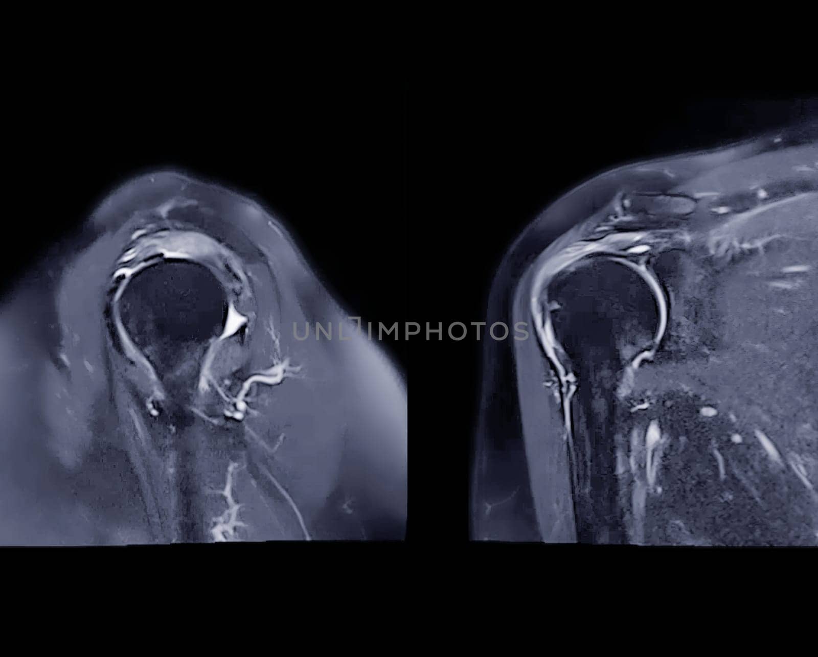
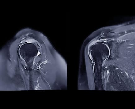
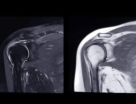
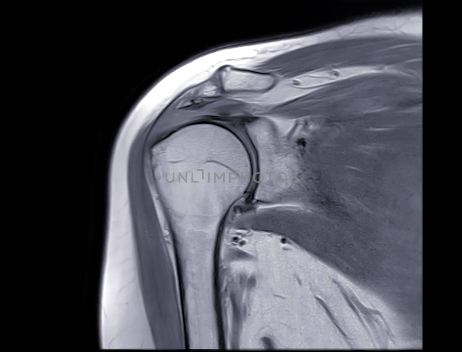
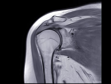
Magnetic Resonance Imaging or MRI of Shoulder Joint Sagittal and Coronal T2 FS for diagnostic shoulder pain.
Stock PhotoUsername
samunellaResolution
3224x2592pxMagnetic Resonance Imaging or MRI of Shoulder Joint Sagittal and Coronal T2 FS for diagnostic shoulder pain.
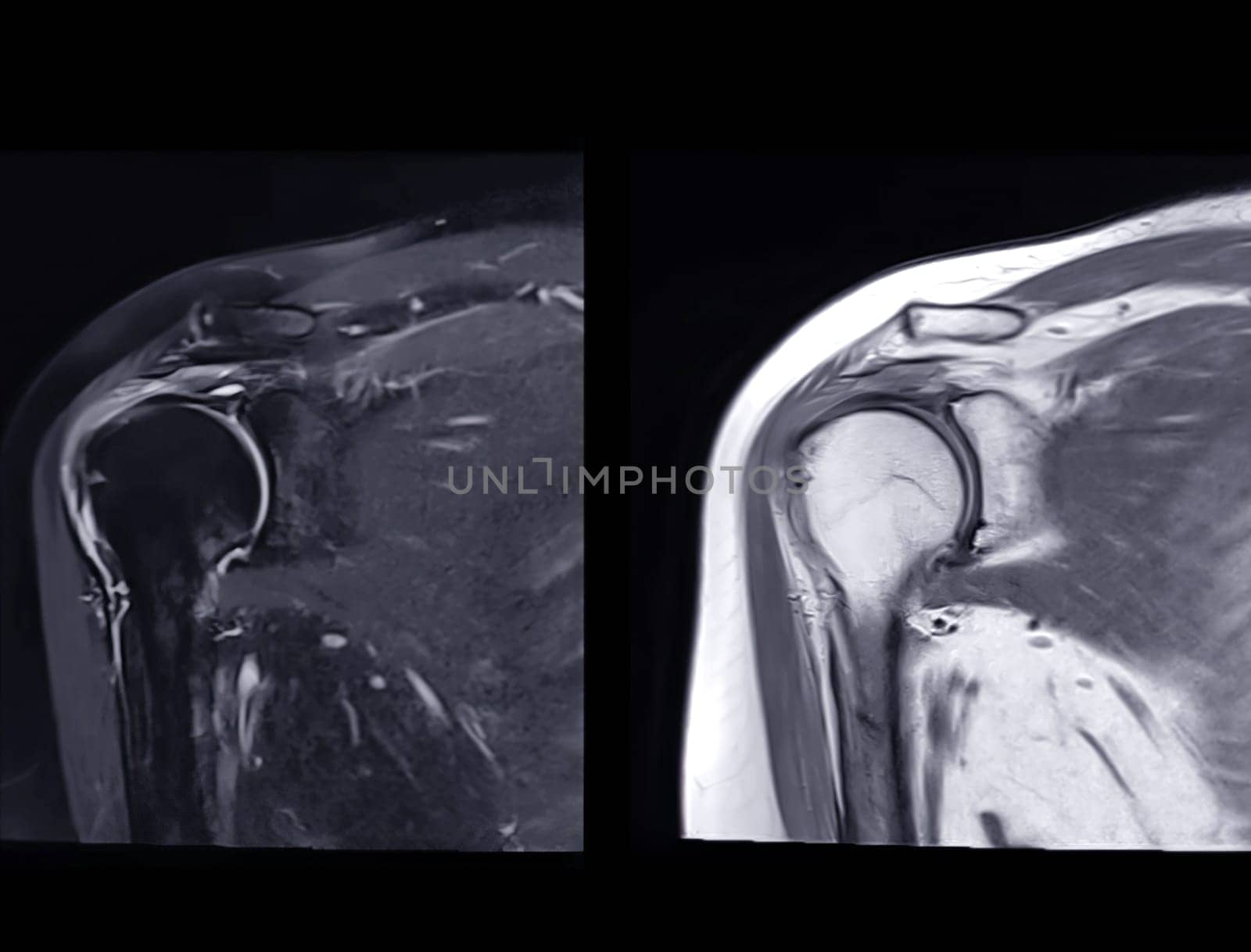
Magnetic Resonance Imaging or MRI of Shoulder Joint Coronal T2 FS and PDW for diagnostic shoulder pain.
Stock PhotoUsername
samunellaResolution
4035x3071pxMagnetic Resonance Imaging or MRI of Shoulder Joint Coronal T2 FS and PDW for diagnostic shoulder pain.
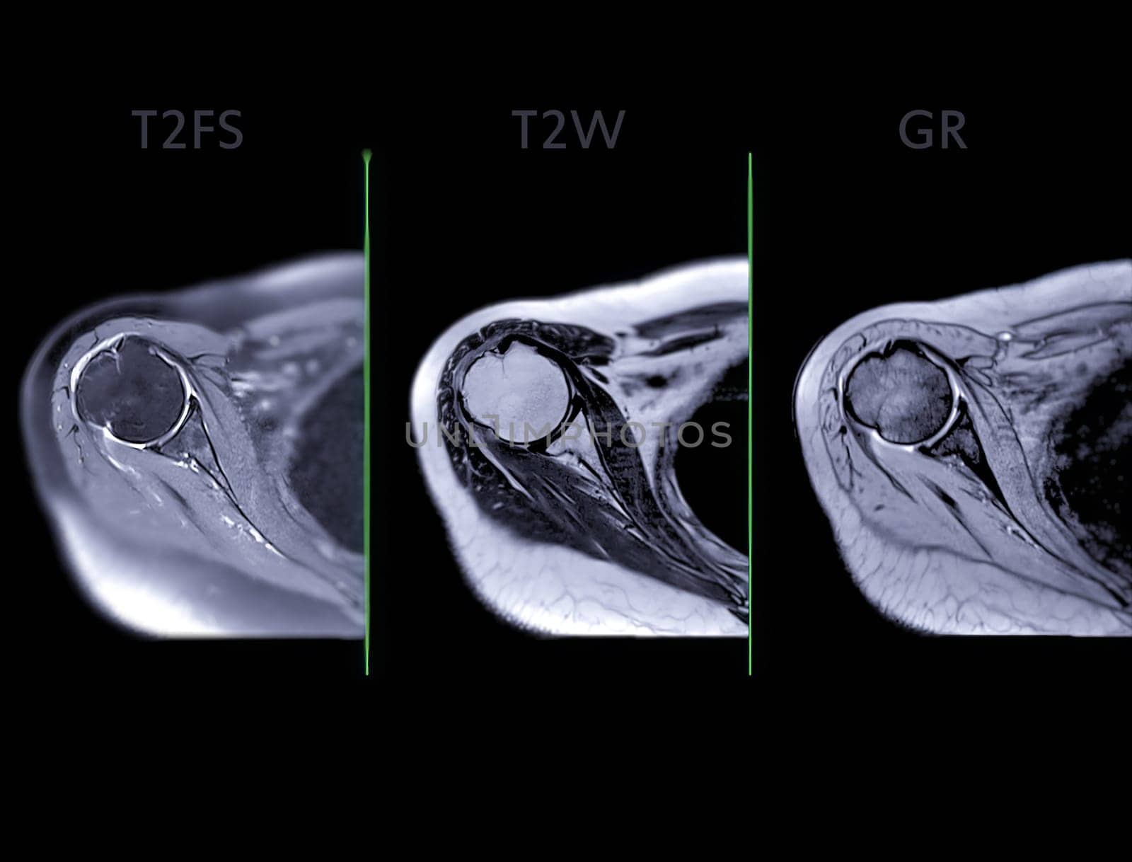
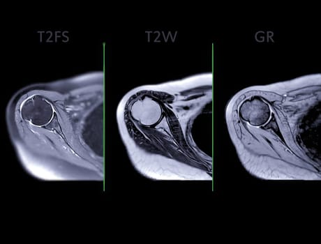
Magnetic Resonance Imaging or MRI of Shoulder Joint Axial T2FS,T2W and mFFE for diagnostic shoulder pain.
Stock PhotoUsername
samunellaResolution
4035x3071pxMagnetic Resonance Imaging or MRI of Shoulder Joint Axial T2FS,T2W and mFFE for diagnostic shoulder pain.
Magnetic Resonance Imaging or MRI of Shoulder Joint Coronal PDW for diagnostic shoulder pain.
Stock PhotoUsername
samunellaResolution
4035x3071pxMagnetic Resonance Imaging or MRI of Shoulder Joint Coronal PDW for diagnostic shoulder pain.
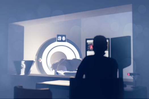
Patient undergoing MRI - Magnetic resonance imaging in Hospital. Medical Equipment and Health Care.3D rendering.
Stock PhotoUsername
samunellaResolution
6000x4000pxPatient undergoing MRI - Magnetic resonance imaging in Hospital. Medical Equipment and Health Care.3D rendering.
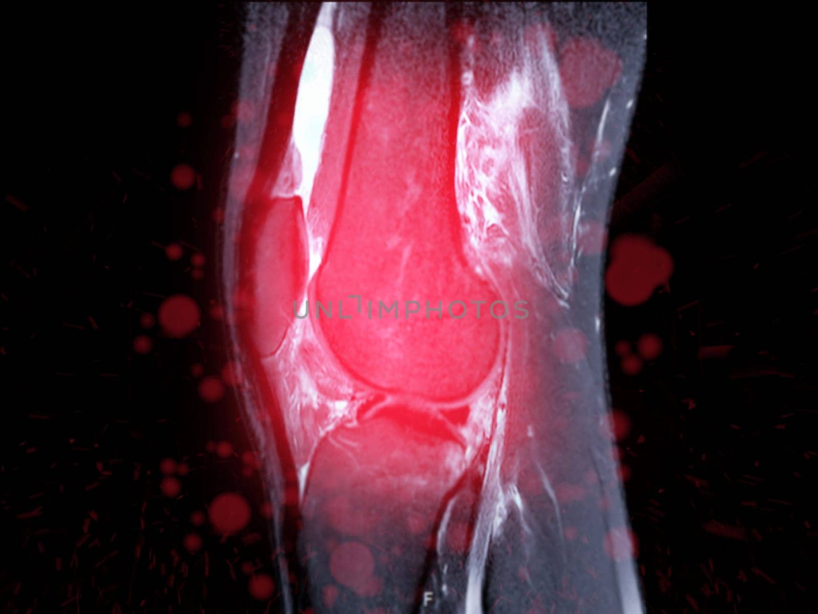
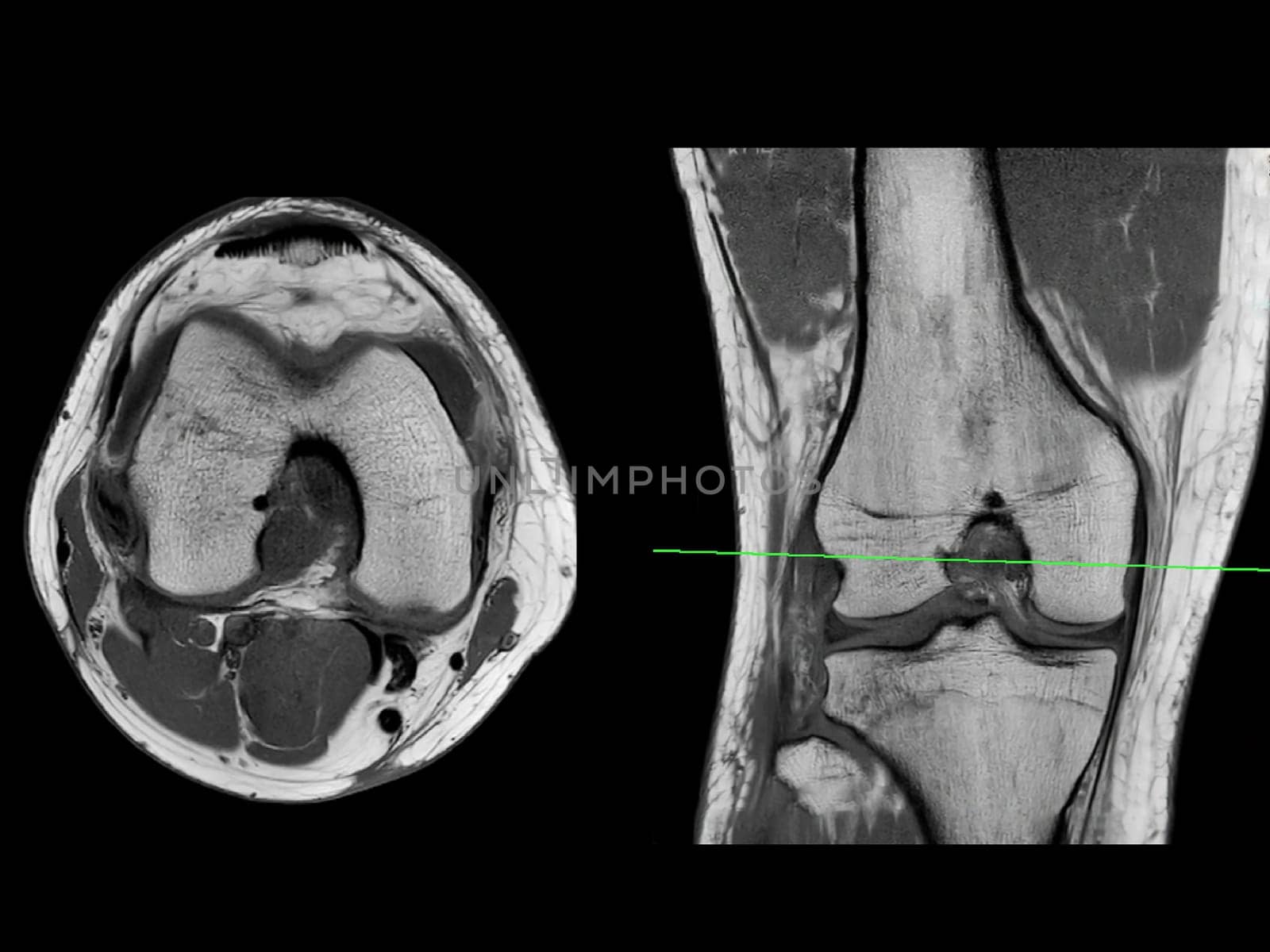
Magnetic resonance imaging or MRI of knee joint Sagittal T2 FS for detect tear or sprain of the anterior cruciate ligament (ACL)
Stock PhotoUsername
samunellaResolution
4000x3000pxMagnetic resonance imaging or MRI of knee joint Sagittal T2 FS for detect tear or sprain of the anterior cruciate ligament (ACL)

Magnetic resonance imaging or MRI of knee joint Sagittal PDW and T2 FS for detect tear or sprain of the anterior cruciate ligament (ACL)
Stock PhotoUsername
samunellaResolution
4000x3000pxMagnetic resonance imaging or MRI of knee joint Sagittal PDW and T2 FS for detect tear or sprain of the anterior cruciate ligament (ACL)

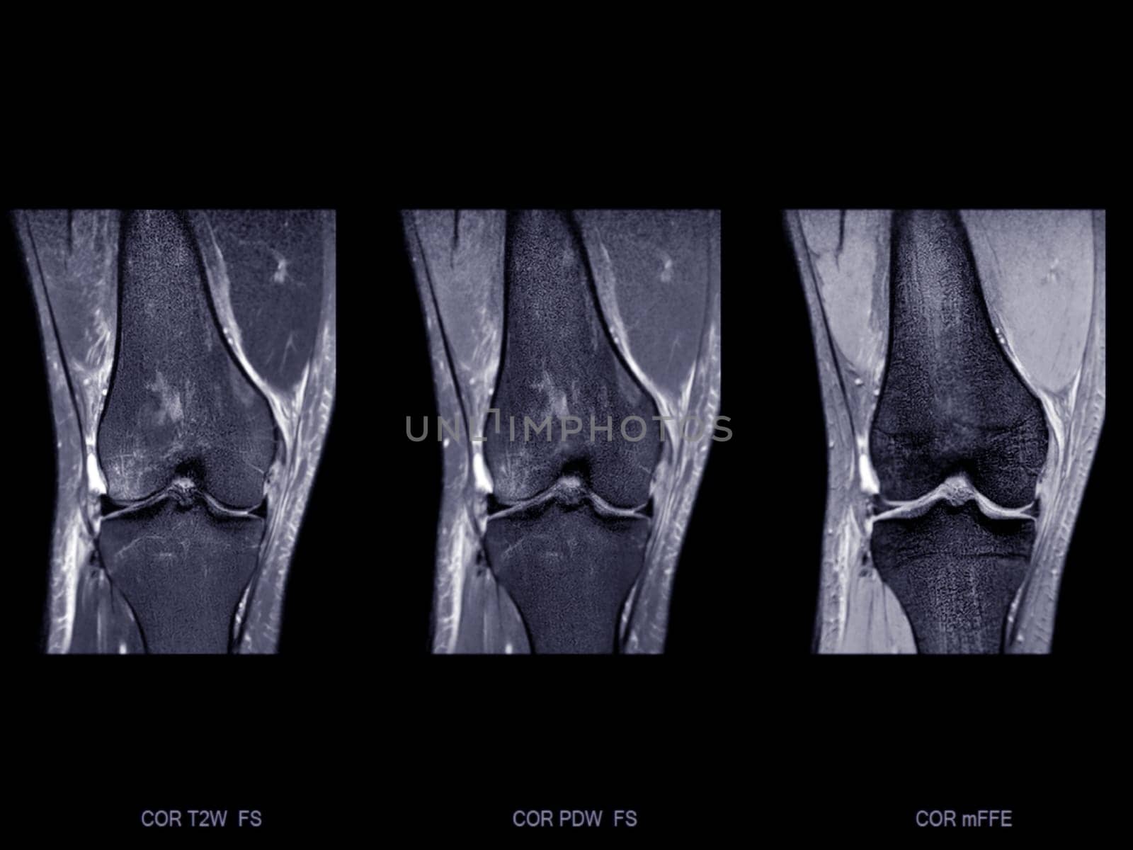
Magnetic resonance imaging or MRI of knee joint Corona; T2 FS , PDW and Gradient for detect tear or sprain of the anterior cruciate ligament (ACL)
Stock PhotoUsername
samunellaResolution
4000x3000pxMagnetic resonance imaging or MRI of knee joint Corona; T2 FS , PDW and Gradient for detect tear or sprain of the anterior cruciate ligament (ACL)
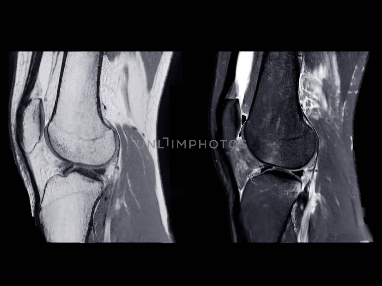
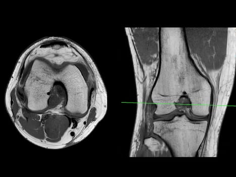
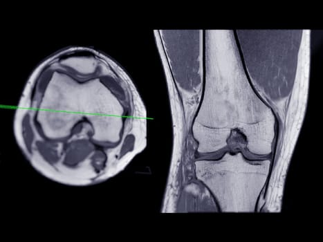
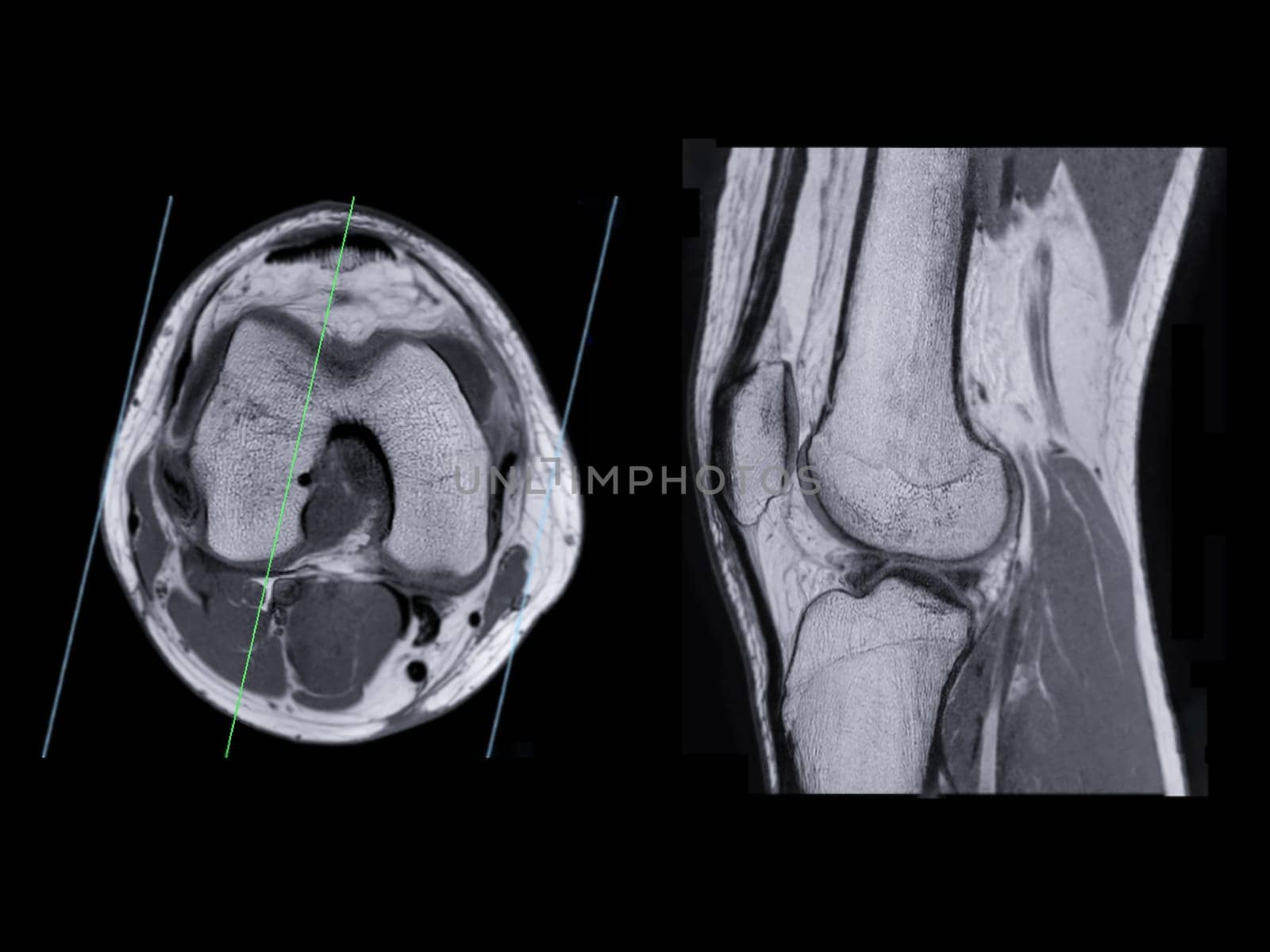
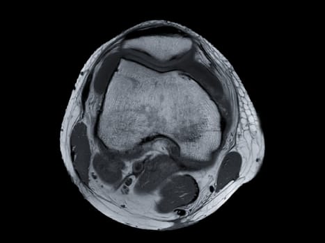
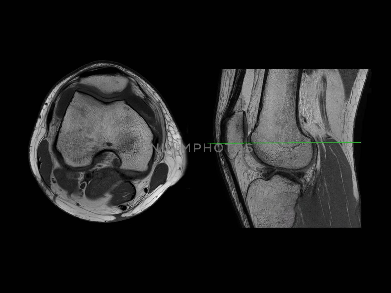
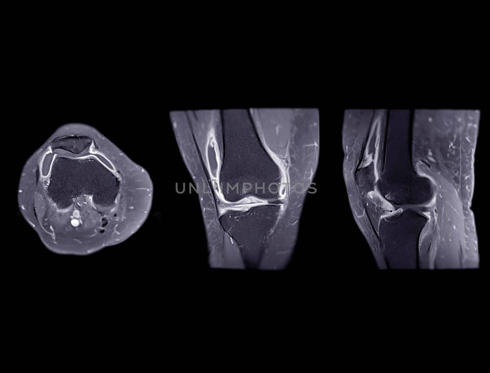
Magnetic resonance imaging or MRI of knee joint Axial T2 and Coronal view for detect tear or sprain of the anterior cruciate ligament (ACL)
Stock PhotoUsername
samunellaResolution
4000x3000pxMagnetic resonance imaging or MRI of knee joint Axial T2 and Coronal view for detect tear or sprain of the anterior cruciate ligament (ACL)
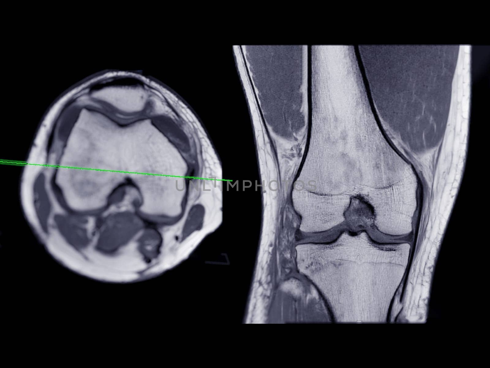
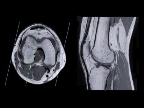
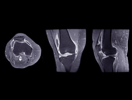
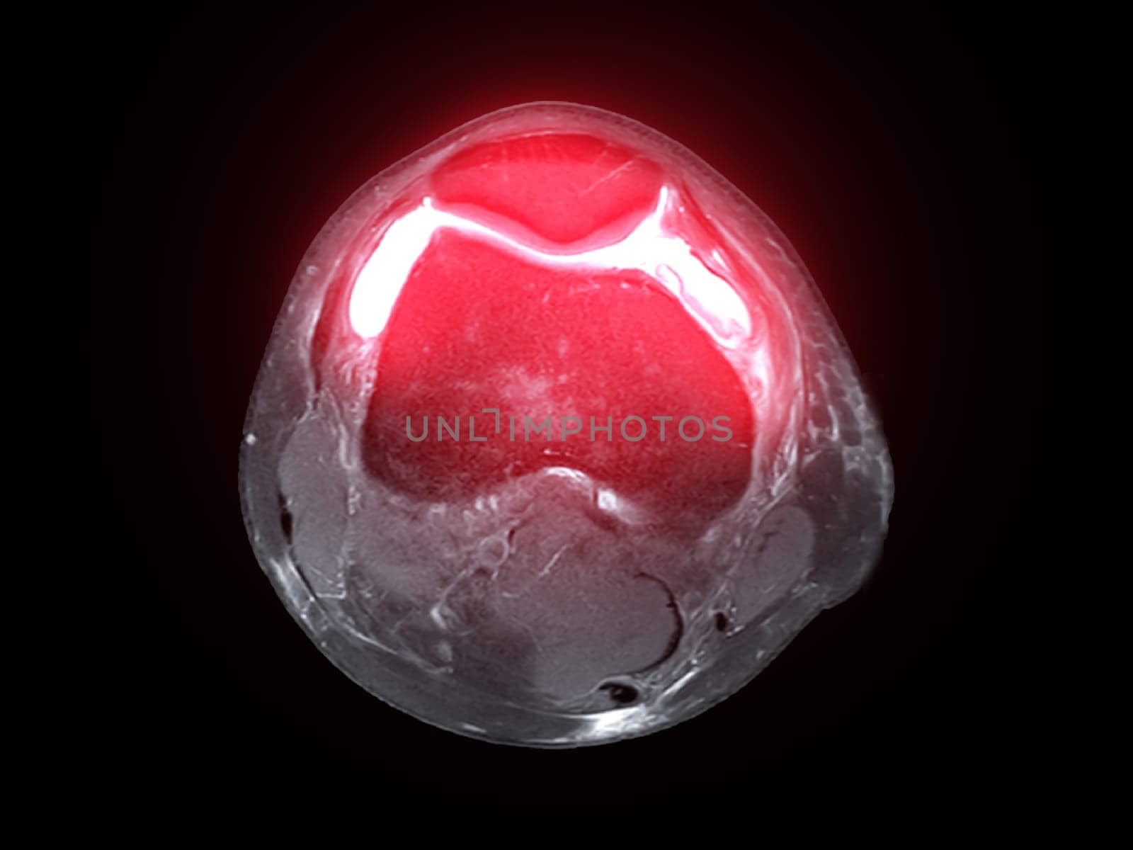
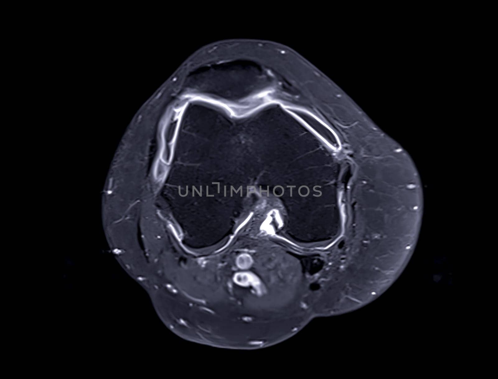
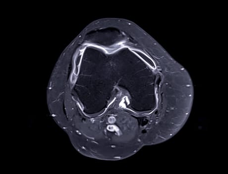
Magnetic resonance imaging or MRI of knee joint Axial T2 FS view for detect tear or sprain of the anterior cruciate ligament (ACL)
Stock PhotoUsername
samunellaResolution
4000x3000pxMagnetic resonance imaging or MRI of knee joint Axial T2 FS view for detect tear or sprain of the anterior cruciate ligament (ACL)
Magnetic resonance imaging or MRI of knee joint Axial and sagittal PDW for detect tear or sprain of the anterior cruciate ligament (ACL)
Stock PhotoUsername
samunellaResolution
4000x3000pxMagnetic resonance imaging or MRI of knee joint Axial and sagittal PDW for detect tear or sprain of the anterior cruciate ligament (ACL)
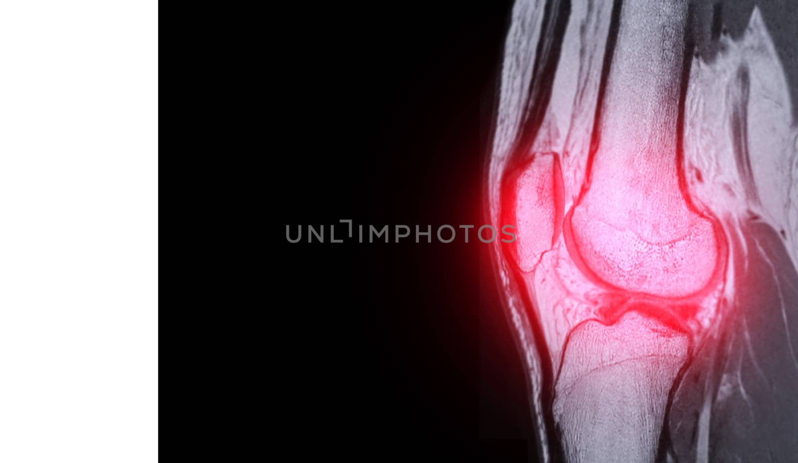
Magnetic resonance imaging or MRI of knee joint Axial view T2 FS with Gadolinium for detect tear or sprain of the anterior cruciate ligament (ACL)
Stock PhotoUsername
samunellaResolution
4035x3071pxMagnetic resonance imaging or MRI of knee joint Axial view T2 FS with Gadolinium for detect tear or sprain of the anterior cruciate ligament (ACL)

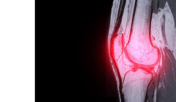
Magnetic resonance imaging or MRI of knee joint Axial view for detect tear or sprain of the anterior cruciate ligament (ACL)
Stock PhotoUsername
samunellaResolution
4000x3000pxMagnetic resonance imaging or MRI of knee joint Axial view for detect tear or sprain of the anterior cruciate ligament (ACL)

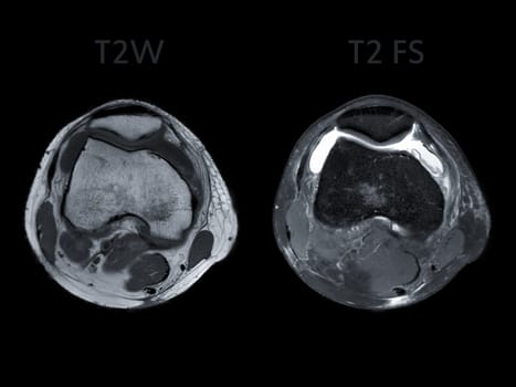
Magnetic resonance imaging or MRI of knee joint Axial view T2 and T2 FS for detect tear or sprain of the anterior cruciate ligament (ACL)
Stock PhotoUsername
samunellaResolution
4000x3000pxMagnetic resonance imaging or MRI of knee joint Axial view T2 and T2 FS for detect tear or sprain of the anterior cruciate ligament (ACL)
Magnetic resonance imaging or MRI of knee joint Axial and sagittal PDW for detect tear or sprain of the anterior cruciate ligament (ACL)
Stock PhotoUsername
samunellaResolution
4000x3000pxMagnetic resonance imaging or MRI of knee joint Axial and sagittal PDW for detect tear or sprain of the anterior cruciate ligament (ACL)

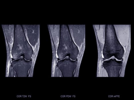
Magnetic resonance imaging or MRI of knee joint Axial ,Coronal and sagittal T2 FS for detect tear or sprain of the anterior cruciate ligament (ACL)
Stock PhotoUsername
samunellaResolution
4035x3071pxMagnetic resonance imaging or MRI of knee joint Axial ,Coronal and sagittal T2 FS for detect tear or sprain of the anterior cruciate ligament (ACL)
Magnetic resonance imaging or MRI of knee joint sagittal PDW for detect tear or sprain of the anterior cruciate ligament (ACL)
Stock PhotoUsername
samunellaResolution
5168x3000pxMagnetic resonance imaging or MRI of knee joint sagittal PDW for detect tear or sprain of the anterior cruciate ligament (ACL)

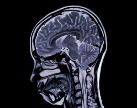
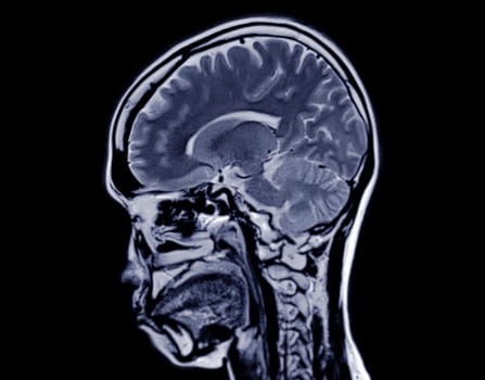
MRI brain scan sagittal plane for detect Brain diseases sush as stroke disease, Brain tumors and Infections.
Stock PhotoUsername
samunellaResolution
3456x2944pxMRI brain scan sagittal plane for detect Brain diseases sush as stroke disease, Brain tumors and Infections.

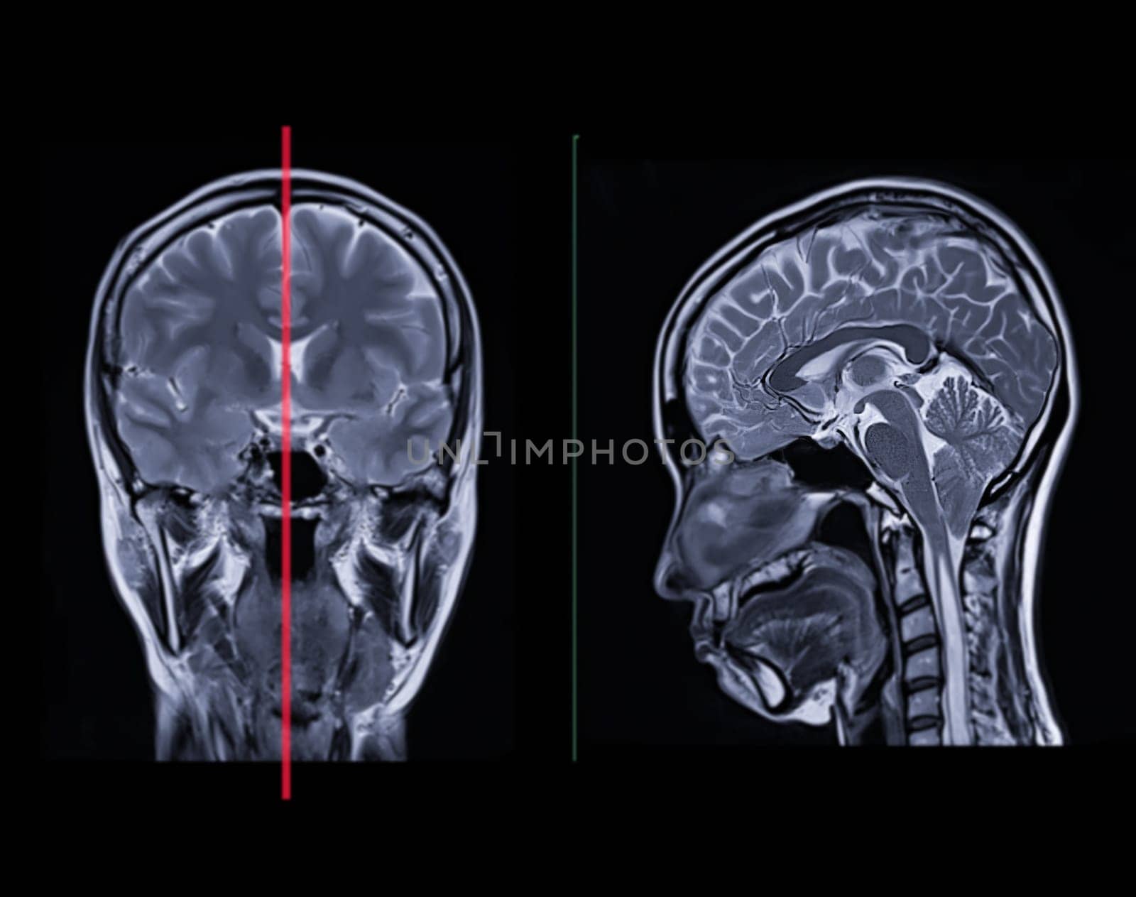
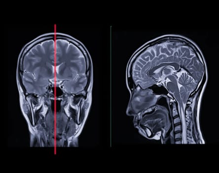
MRI brain scan Compare Coronal and sagittal plane for detect Brain diseases sush as stroke disease, Brain tumors and Infections.
Stock PhotoUsername
samunellaResolution
4035x3071pxMRI brain scan Compare Coronal and sagittal plane for detect Brain diseases sush as stroke disease, Brain tumors and Infections.
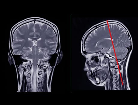

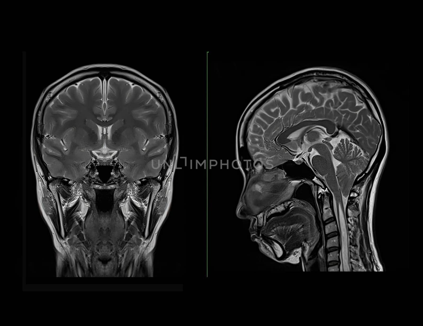
MRI brain scan Compare Coronal and sagittal plane for detect Brain diseases sush as stroke disease, Brain tumors and Infections.
Stock PhotoUsername
samunellaResolution
3303x2695pxMRI brain scan Compare Coronal and sagittal plane for detect Brain diseases sush as stroke disease, Brain tumors and Infections.
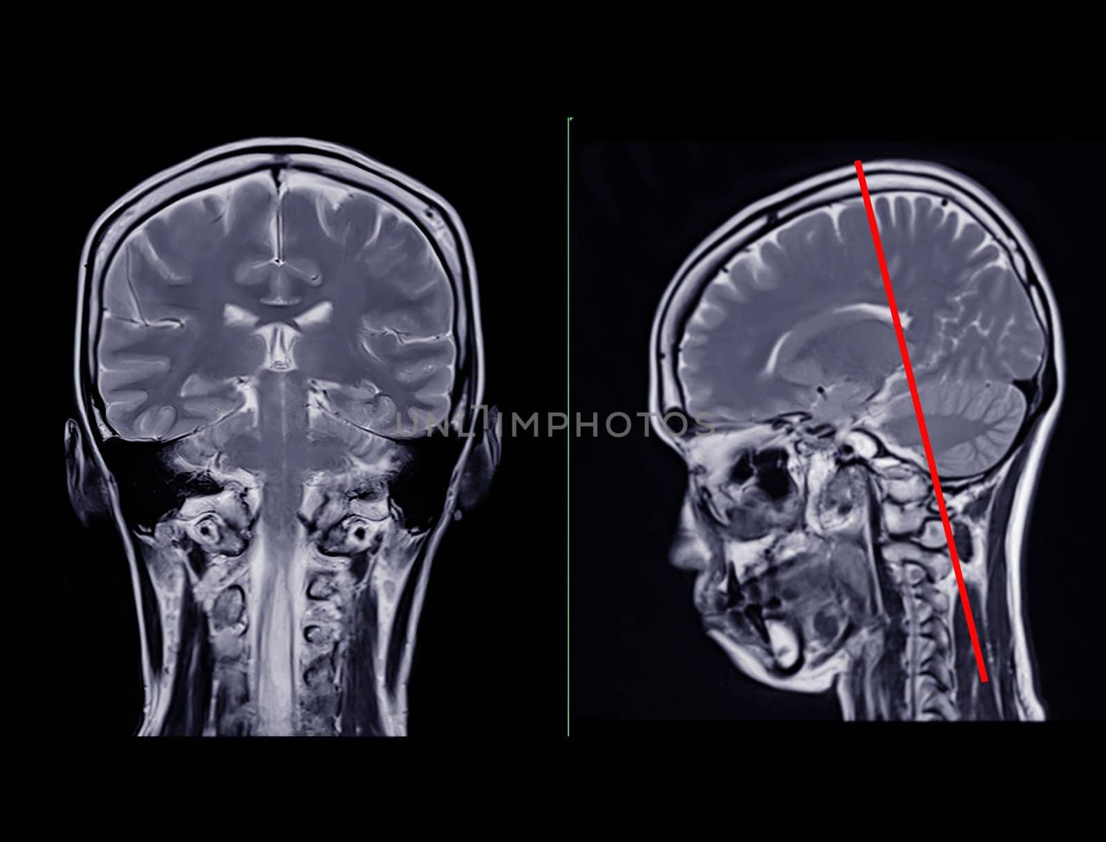
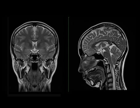
MRI brain scan Compare Coronal and sagittal plane for detect Brain diseases sush as stroke disease, Brain tumors and Infections.
Stock PhotoUsername
samunellaResolution
3614x2852pxMRI brain scan Compare Coronal and sagittal plane for detect Brain diseases sush as stroke disease, Brain tumors and Infections.
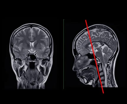
MRI brain scan Compare Coronal and sagittal plane for detect Brain diseases sush as stroke disease, Brain tumors and Infections.
Stock PhotoUsername
samunellaResolution
3568x2752pxMRI brain scan Compare Coronal and sagittal plane for detect Brain diseases sush as stroke disease, Brain tumors and Infections.
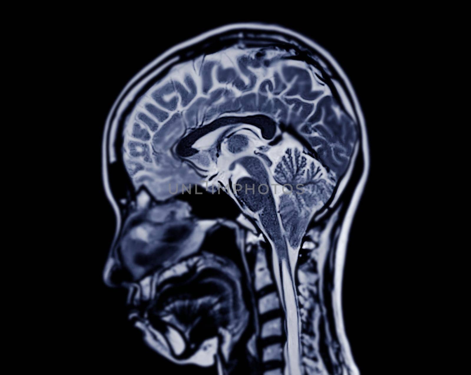
MRI brain scan sagittal plane for detect Brain diseases sush as stroke disease, Brain tumors and Infections.
Stock PhotoUsername
samunellaResolution
3649x2911pxMRI brain scan sagittal plane for detect Brain diseases sush as stroke disease, Brain tumors and Infections.


MRI brain scan sagittal plane for detect Brain diseases sush as stroke disease, Brain tumors and Infections.
Stock PhotoUsername
samunellaResolution
3283x2592pxMRI brain scan sagittal plane for detect Brain diseases sush as stroke disease, Brain tumors and Infections.
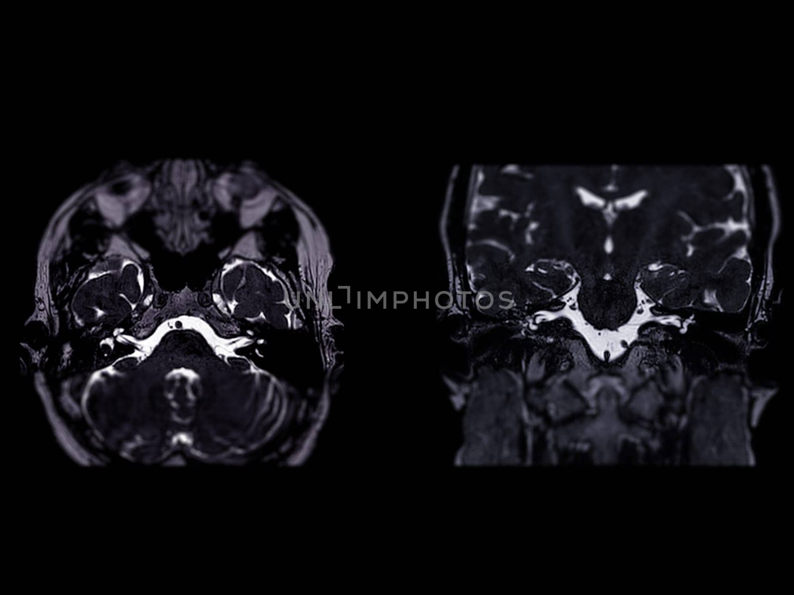
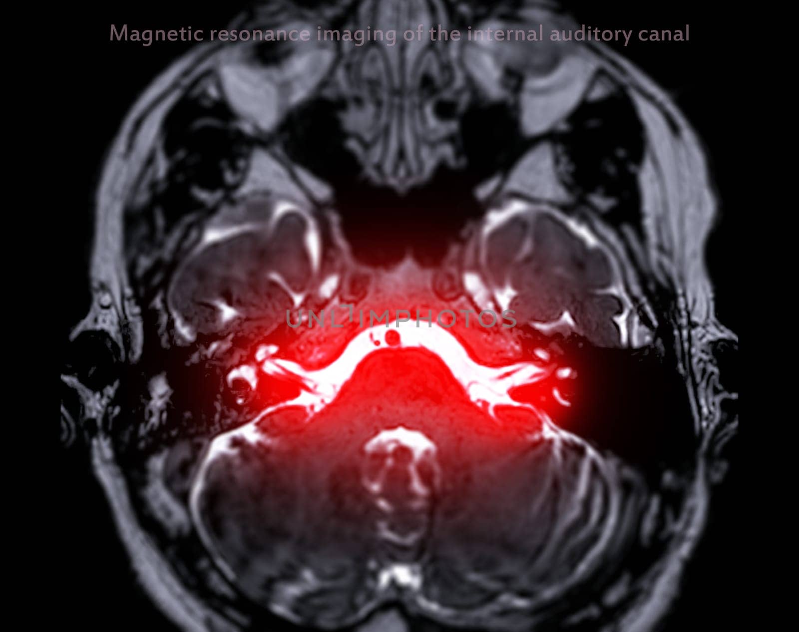
MRI Brain scan with the internal auditory canal (IAC) axial and Coronal view.
Stock PhotoUsername
samunellaResolution
4000x3000pxMRI Brain scan with the internal auditory canal (IAC) axial and Coronal view.
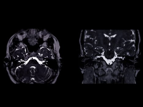
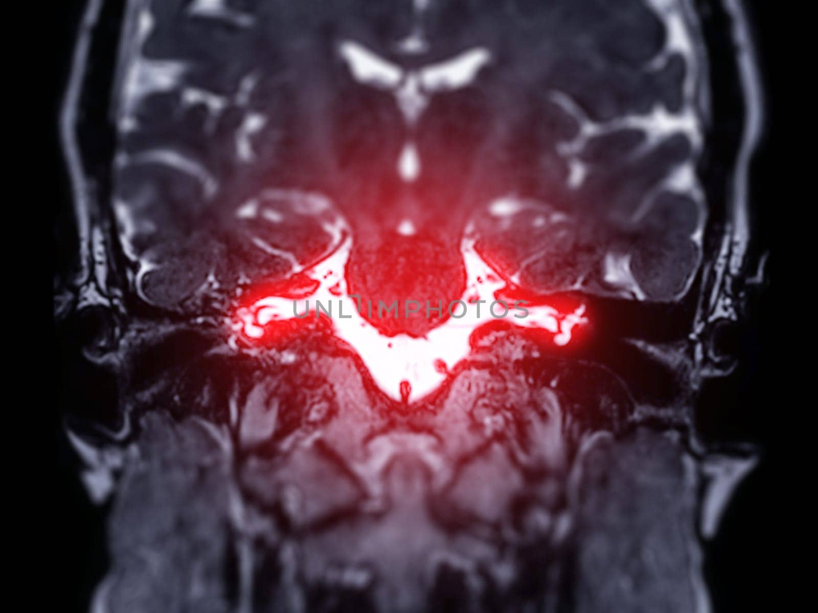
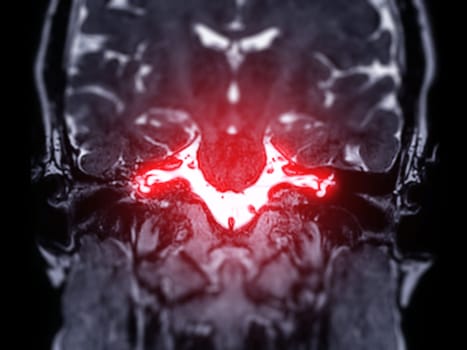
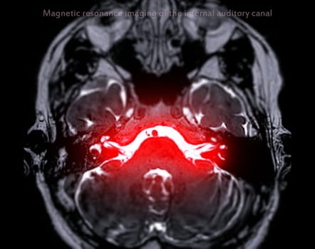
MRI Brain scan with the internal auditory canal (IAC) Coronal view.
Stock PhotoUsername
samunellaResolution
4000x3000pxMRI Brain scan with the internal auditory canal (IAC) Coronal view.
MRI Brain scan with the internal auditory canal (IAC) axial view.
Stock PhotoUsername
samunellaResolution
3044x2408pxMRI Brain scan with the internal auditory canal (IAC) axial view.
Magnetic resonance imaging or MRI of knee joint Axial T2 FS view for detect tear or sprain of the anterior cruciate ligament (ACL)
Stock PhotoUsername
samunellaResolution
5190x3731pxMagnetic resonance imaging or MRI of knee joint Axial T2 FS view for detect tear or sprain of the anterior cruciate ligament (ACL)

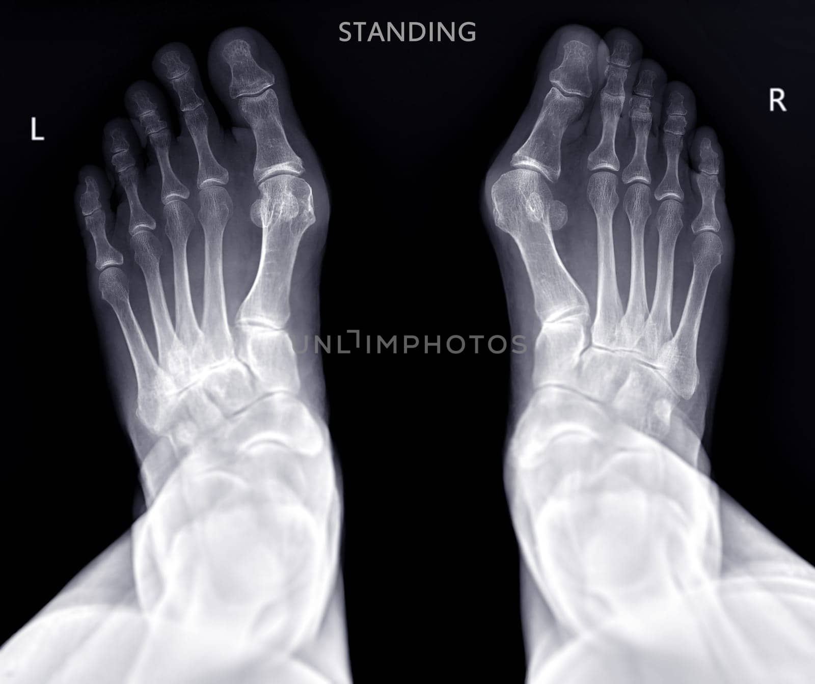
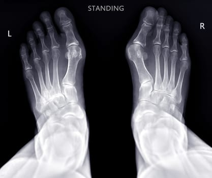
Foot x-ray image AP standing view isolated on black background.
Stock PhotoUsername
samunellaResolution
4226x3546pxFoot x-ray image AP standing view isolated on black background.

MRI brain scan sagittal plane for detect Brain diseases sush as stroke disease, Brain tumors and Infections.
Stock PhotoUsername
samunellaResolution
3506x2744pxMRI brain scan sagittal plane for detect Brain diseases sush as stroke disease, Brain tumors and Infections.
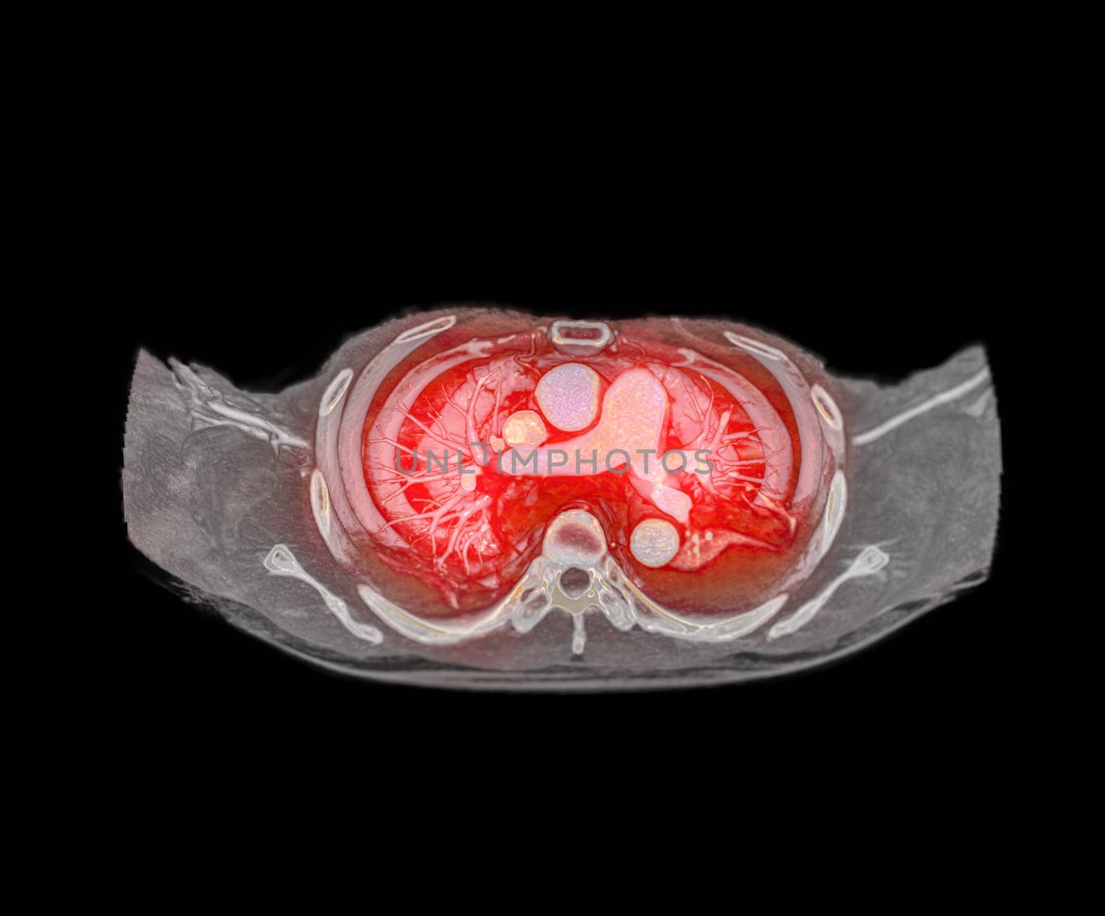
CTA pulmonary arteries 3D rendering showing branch of pulmonary artery
Stock PhotoUsername
samunellaResolution
4038x3347pxCTA pulmonary arteries 3D rendering showing branch of pulmonary artery

CTA pulmonary arteries 3D rendering showing branch of pulmonary artery
Stock PhotoUsername
samunellaResolution
4038x3347pxCTA pulmonary arteries 3D rendering showing branch of pulmonary artery

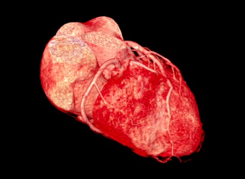
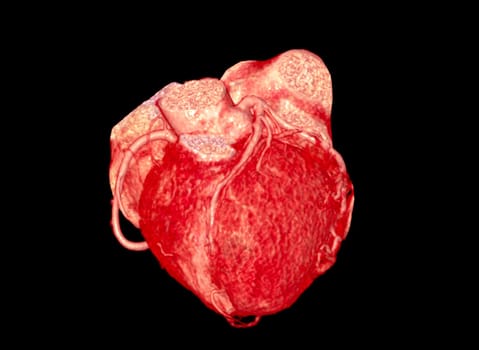
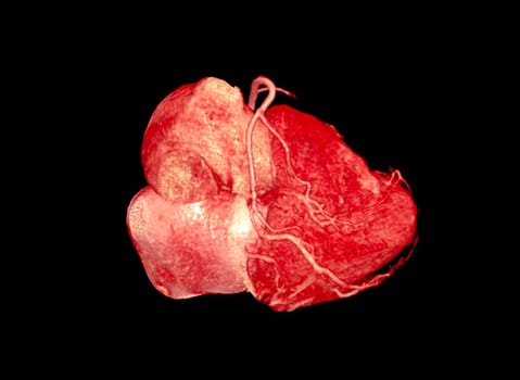
CTA Coronary artery 3D rendering image.
Stock PhotoUsername
samunellaResolution
6900x5052pxCTA Coronary artery 3D rendering image.

CTA Coronary artery 3D rendering image.
Stock PhotoUsername
samunellaResolution
6900x5052pxCTA Coronary artery 3D rendering image.

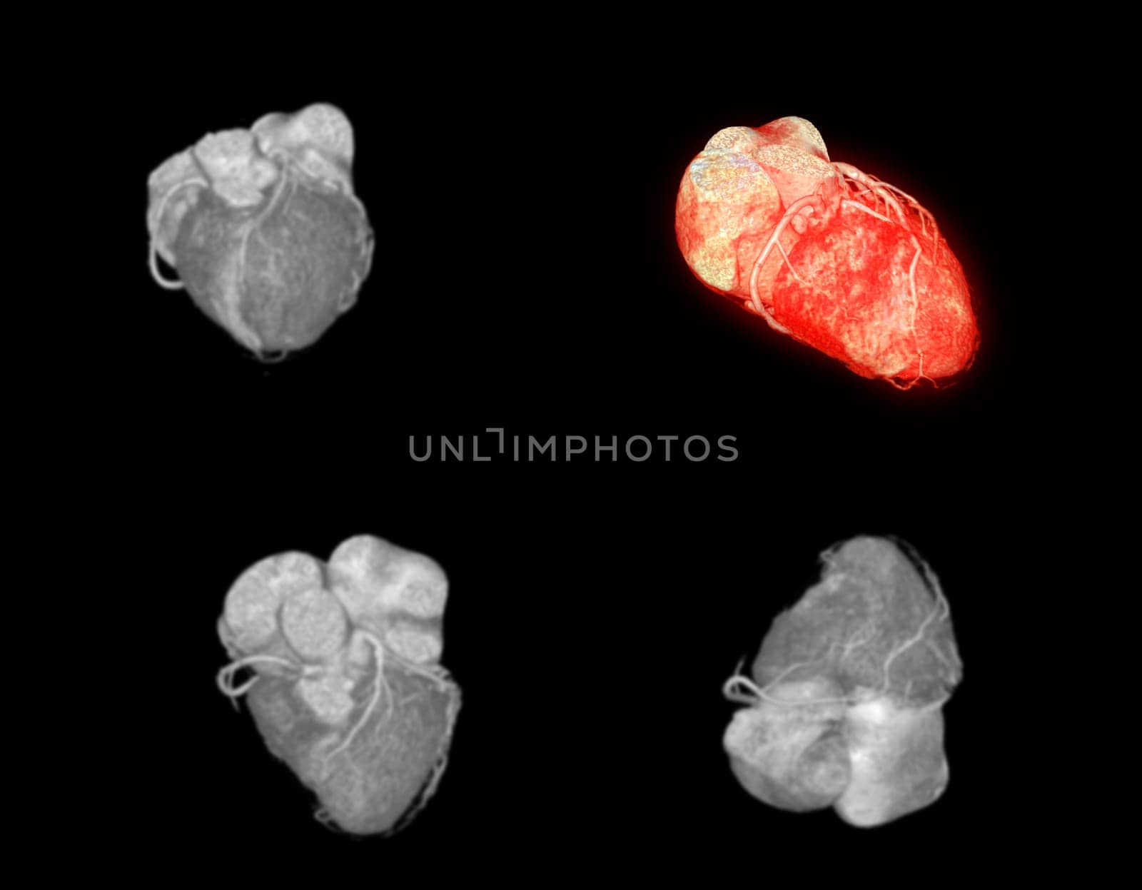
CTA Coronary artery 3D rendering image.
Stock PhotoUsername
samunellaResolution
6900x5052pxCTA Coronary artery 3D rendering image.
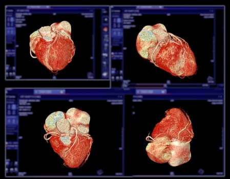
CTA Coronary artery 3D rendering image.
Stock PhotoUsername
samunellaResolution
6000x4679pxCTA Coronary artery 3D rendering image.


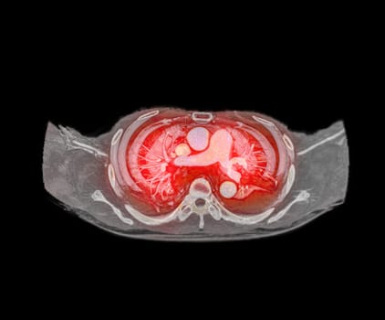
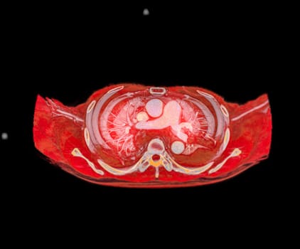
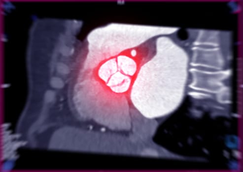
CTA of the aorta showing aortic valve.
Stock PhotoUsername
samunellaResolution
6216x4415pxCTA of the aorta showing aortic valve.

CTA Coronary artery 3D rendering image.
Stock PhotoUsername
samunellaResolution
6000x4679pxCTA Coronary artery 3D rendering image.
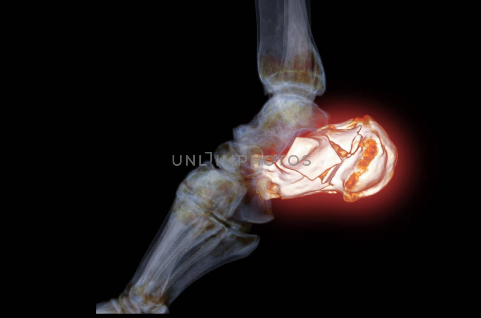
CT Scan ankle joint with 3d rendering of calcaneus bone showing Calcaneus (Heel Bone) Fractures.
Stock PhotoUsername
samunellaResolution
4260x2820pxCT Scan ankle joint with 3d rendering of calcaneus bone showing Calcaneus (Heel Bone) Fractures.

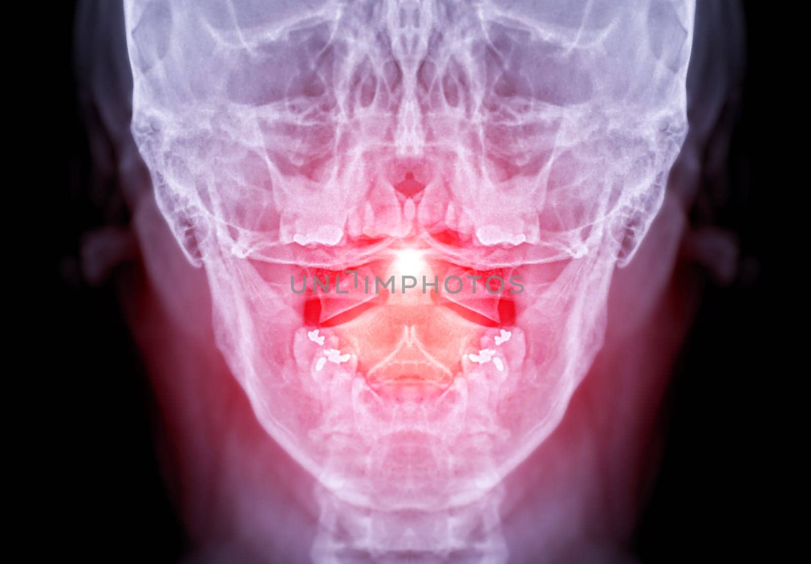
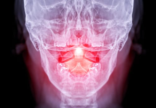
X-ray C-spine or x-ray image of Cervical spine open mount view for fracture of cervical vertebra 2nd ( axis ).
Stock PhotoUsername
samunellaResolution
3356x2336pxX-ray C-spine or x-ray image of Cervical spine open mount view for fracture of cervical vertebra 2nd ( axis ).
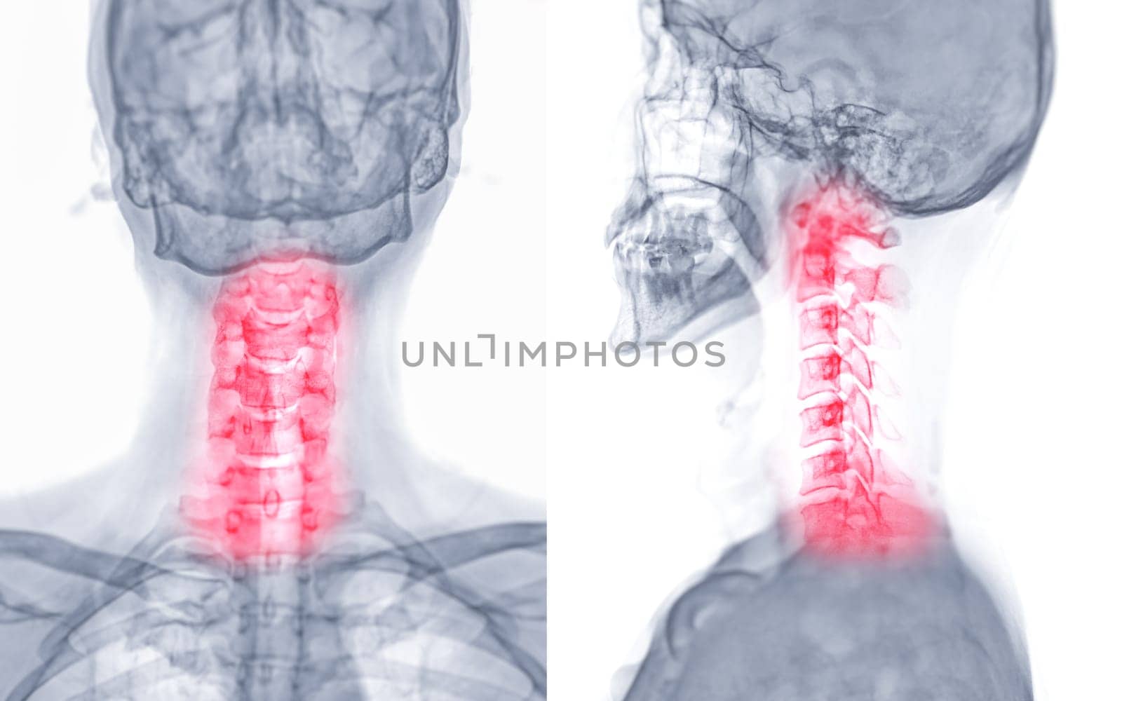
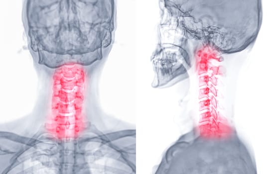
X-ray C-spine or x-ray image of Cervical spine AP and Lateral view for diagnostic intervertebral disc herniation ,Spondylosis and fracture.
Stock PhotoUsername
samunellaResolution
7600x4751pxX-ray C-spine or x-ray image of Cervical spine AP and Lateral view for diagnostic intervertebral disc herniation ,Spondylosis and fracture.