- Filter By:
-
-
Stock photos and images of username:samunella
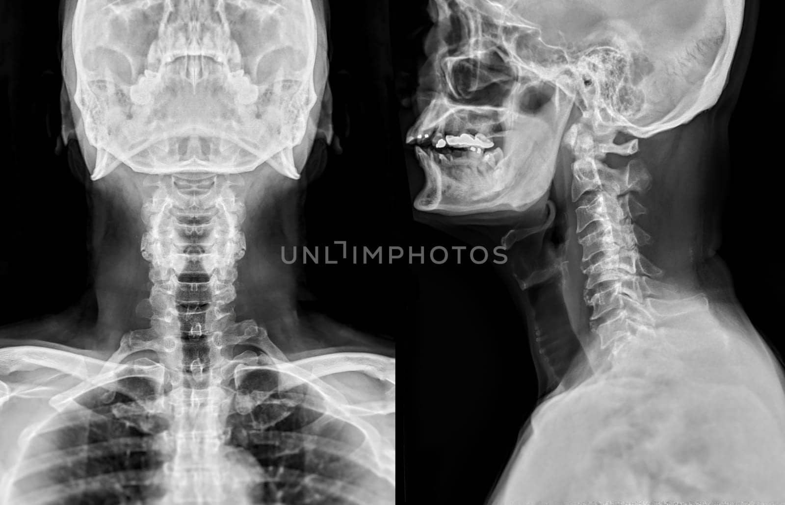
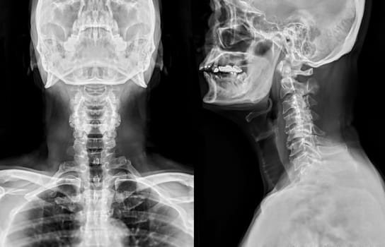
X-ray C-spine or x-ray image of Cervical spine AP and Lateral view for diagnostic intervertebral disc herniation ,Spondylosis and fracture.
Stock PhotoUsername
samunellaResolution
6623x4265pxX-ray C-spine or x-ray image of Cervical spine AP and Lateral view for diagnostic intervertebral disc herniation ,Spondylosis and fracture.
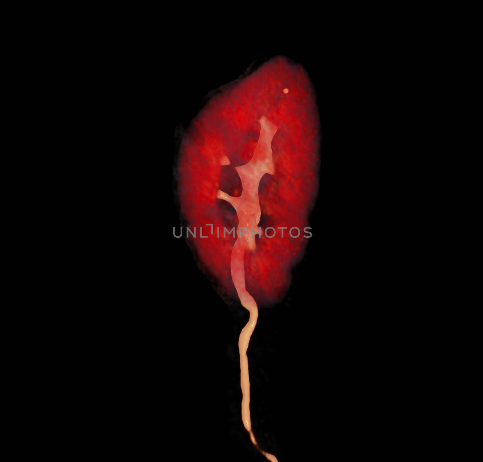
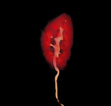
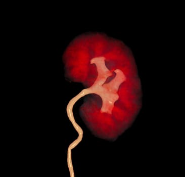
CTA Renal artery showing kidney 3D rendering image.
Stock PhotoUsername
samunellaResolution
3408x3260pxCTA Renal artery showing kidney 3D rendering image.
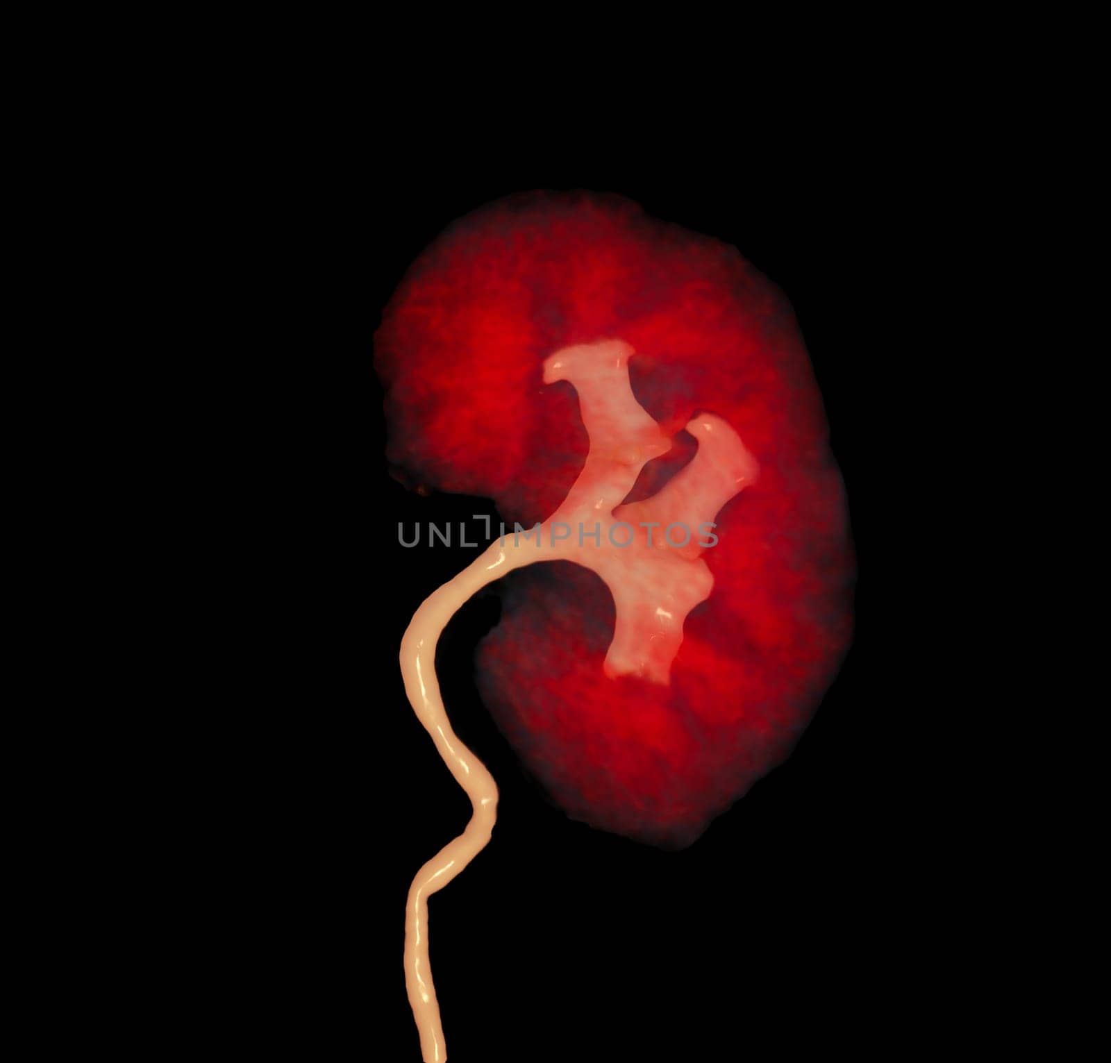
CTA Renal artery showing kidney 3D rendering image.
Stock PhotoUsername
samunellaResolution
3408x3260pxCTA Renal artery showing kidney 3D rendering image.

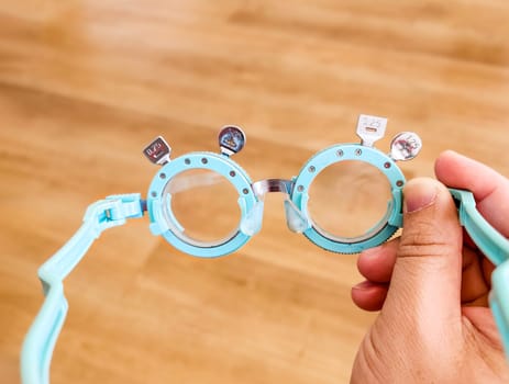
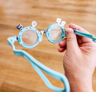
Optometrist . Eye test frame. Vision test. Check Eyesight. Diopter with scale of measurement. Examination of the eyes at eyeglass shop.
Stock PhotoUsername
samunellaResolution
4660x3528pxOptometrist . Eye test frame. Vision test. Check Eyesight. Diopter with scale of measurement. Examination of the eyes at eyeglass shop.

Optometrist . Eye test frame. Vision test. Check Eyesight. Diopter with scale of measurement. Examination of the eyes at eyeglass shop.
Stock PhotoUsername
samunellaResolution
4500x4304pxOptometrist . Eye test frame. Vision test. Check Eyesight. Diopter with scale of measurement. Examination of the eyes at eyeglass shop.

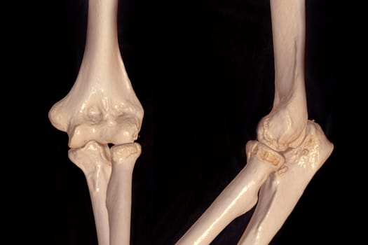
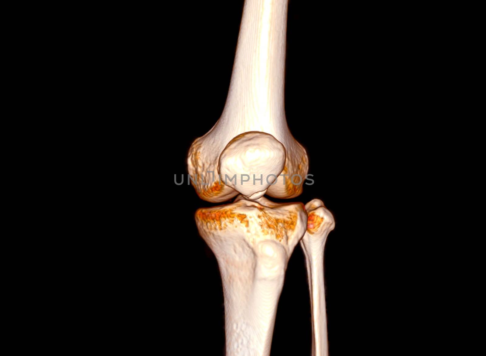
CT scan of elbow joint 3d rendering .
Stock PhotoUsername
samunellaResolution
6000x4000pxCT scan of elbow joint 3d rendering .

Old wood plank or old panels wood texture background.
Stock PhotoUsername
samunellaResolution
4476x3028pxOld wood plank or old panels wood texture background.

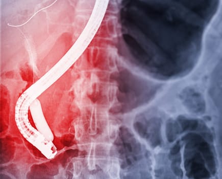
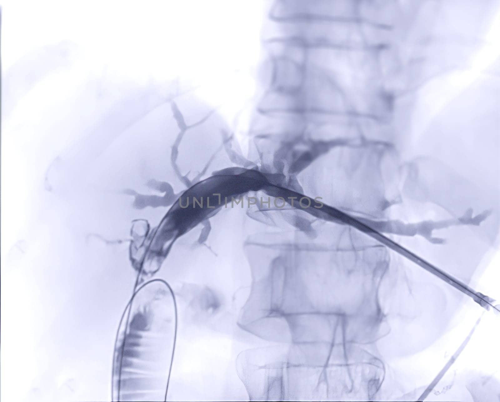
X-ray image of endoscopic after Doctor doing ERCP and laparoscopic cholecystectomy inside modern operating room.
Stock PhotoUsername
samunellaResolution
3794x3052pxX-ray image of endoscopic after Doctor doing ERCP and laparoscopic cholecystectomy inside modern operating room.

X-ray image of endoscopic after Doctor doing ERCP and laparoscopic cholecystectomy inside modern operating room.
Stock PhotoUsername
samunellaResolution
3794x3052pxX-ray image of endoscopic after Doctor doing ERCP and laparoscopic cholecystectomy inside modern operating room.
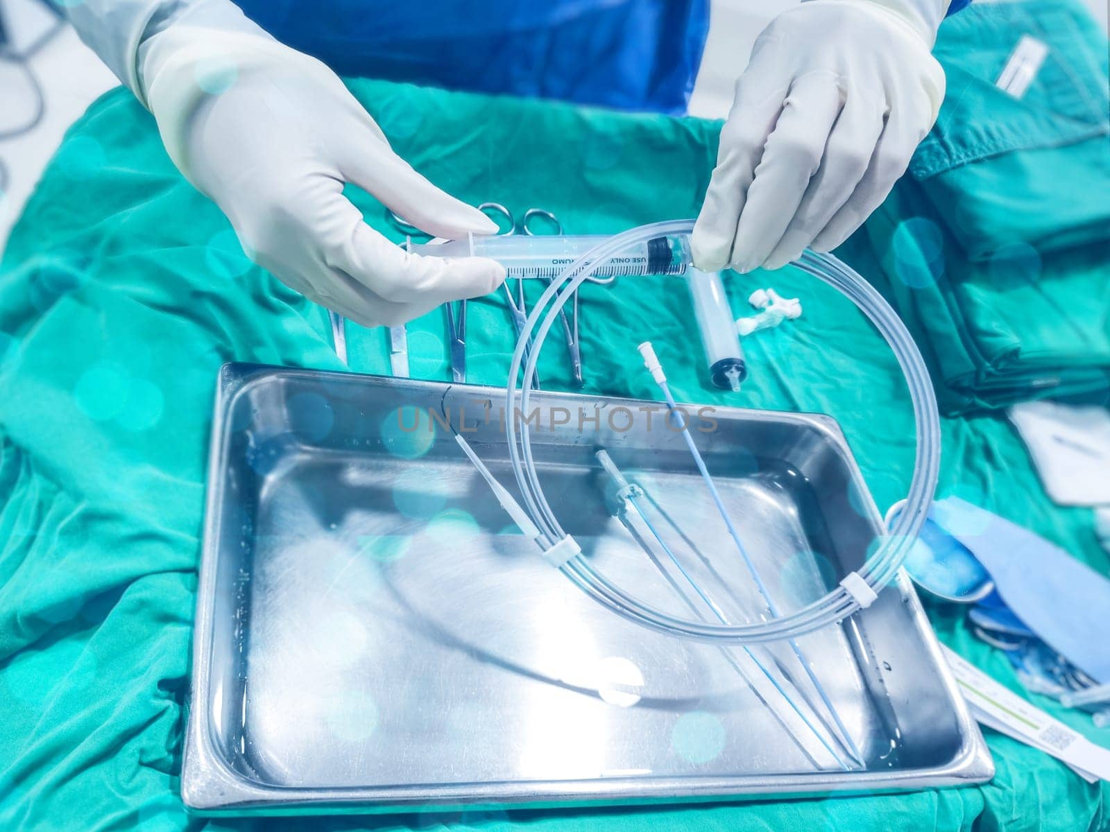
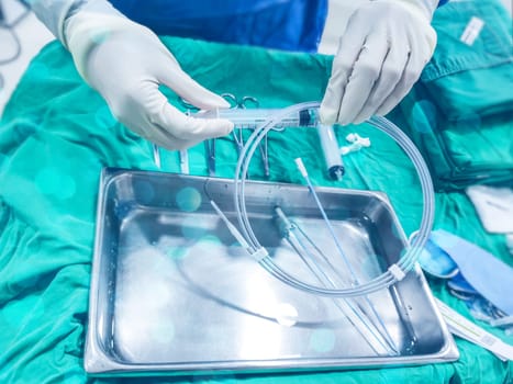
The nurse prepare guidewire for Medical material for surgical intervention packaging and sterile
Stock PhotoUsername
samunellaResolution
4608x3456pxThe nurse prepare guidewire for Medical material for surgical intervention packaging and sterile
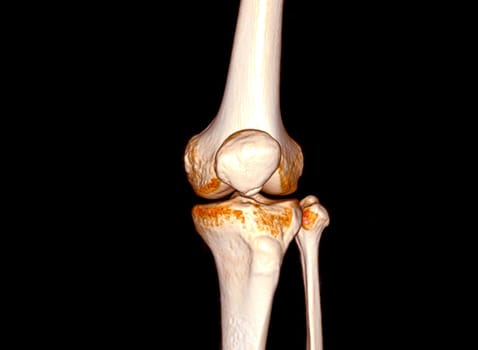
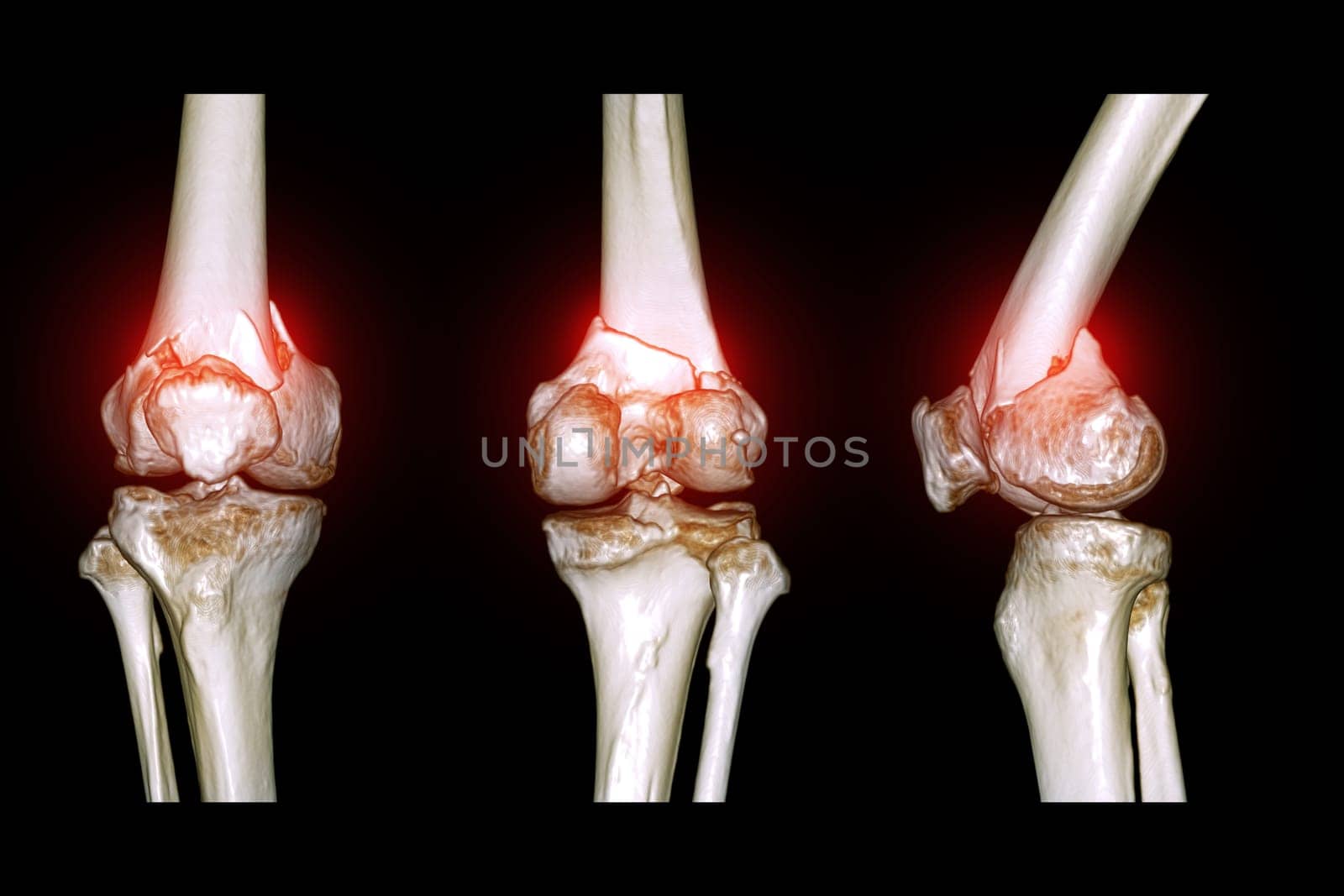
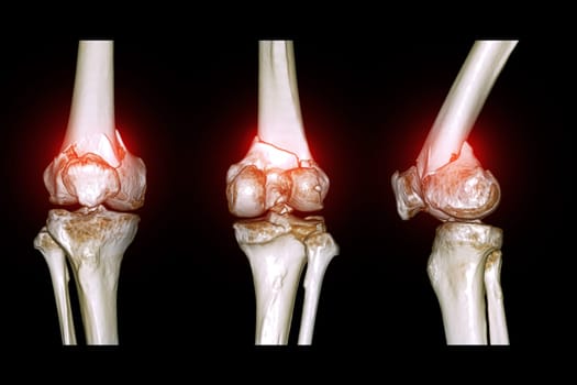
CT Scan of Knee joint 3D rendering .
Stock PhotoUsername
samunellaResolution
4128x3024pxCT Scan of Knee joint 3D rendering .

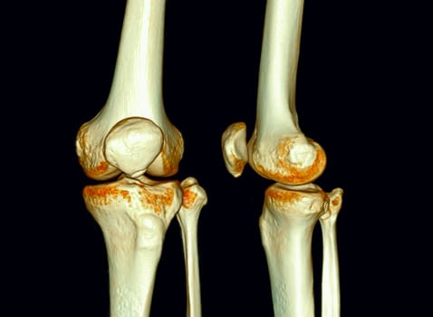
CT Scan of Knee joint 3D rendering .
Stock PhotoUsername
samunellaResolution
4128x3024pxCT Scan of Knee joint 3D rendering .
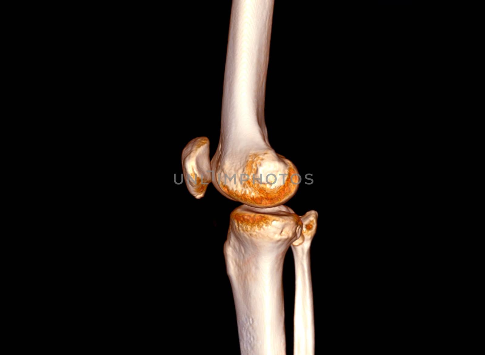
CT Scan of Knee joint 3D rendering .
Stock PhotoUsername
samunellaResolution
4128x3024pxCT Scan of Knee joint 3D rendering .


Woman Doctor with protective workwear holding Vaccine and syringe on CT Scan room background. Female doctor with face mask gloves in Hospital, preparing vaccine.
Stock PhotoUsername
samunellaResolution
4608x3456pxWoman Doctor with protective workwear holding Vaccine and syringe on CT Scan room background. Female doctor with face mask gloves in Hospital, preparing vaccine.


Woman Doctor with protective workwear holding Vaccine and syringe on CT Scan room background. Female doctor with face mask gloves in Hospital, preparing vaccine.
Stock PhotoUsername
samunellaResolution
4608x3073pxWoman Doctor with protective workwear holding Vaccine and syringe on CT Scan room background. Female doctor with face mask gloves in Hospital, preparing vaccine.

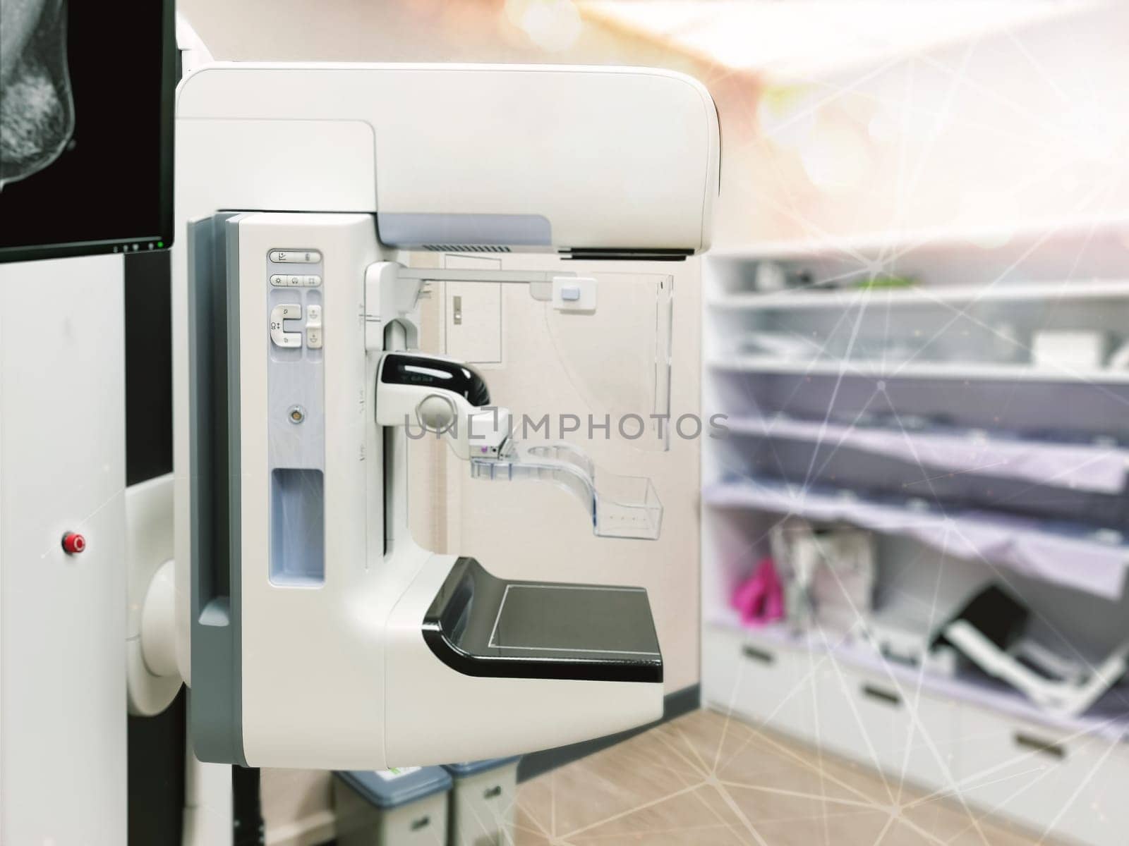
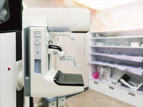
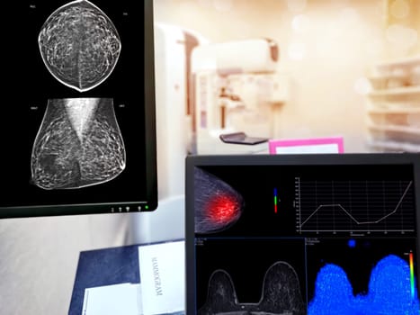
Mammography machine with monitor for breast screening device on mammogram room background.
Stock PhotoUsername
samunellaResolution
4608x3456pxMammography machine with monitor for breast screening device on mammogram room background.

Mammography machine with monitor for breast screening device on mammogram room background.
Stock PhotoUsername
samunellaResolution
4608x3456pxMammography machine with monitor for breast screening device on mammogram room background.
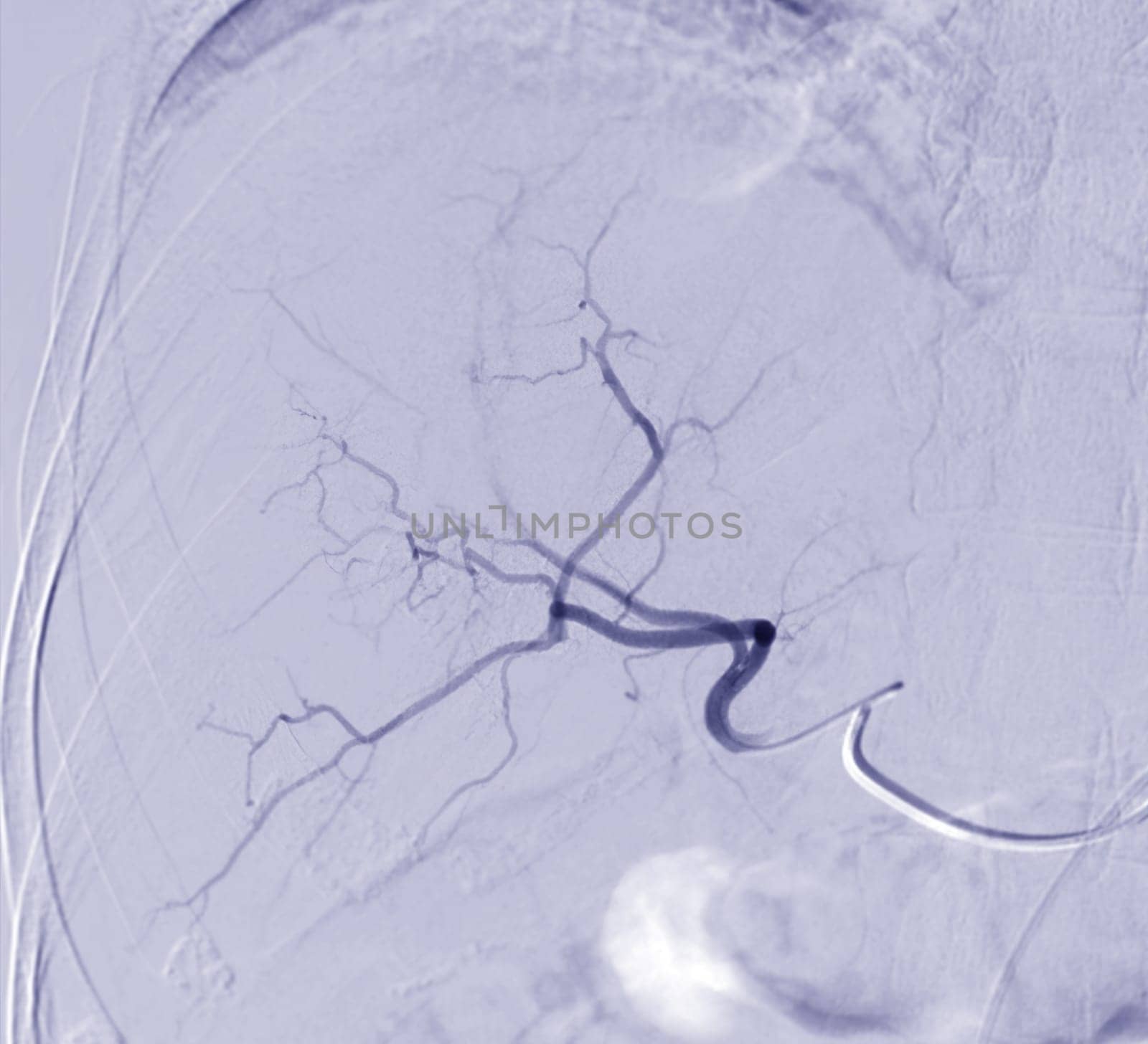
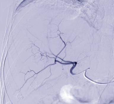
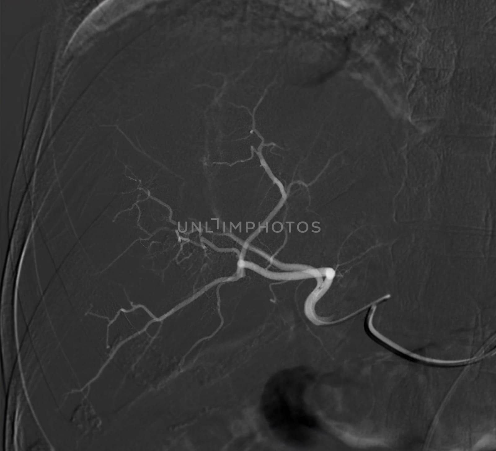
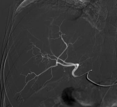
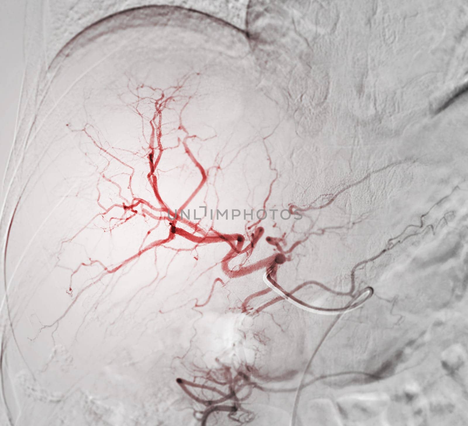
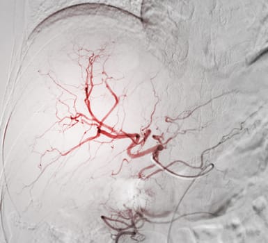
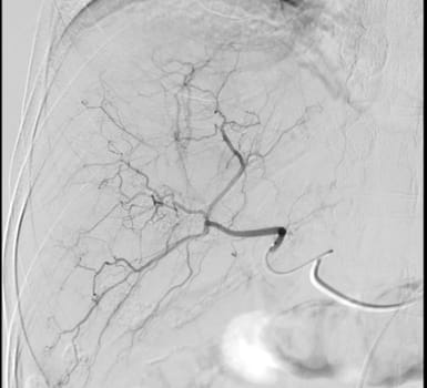
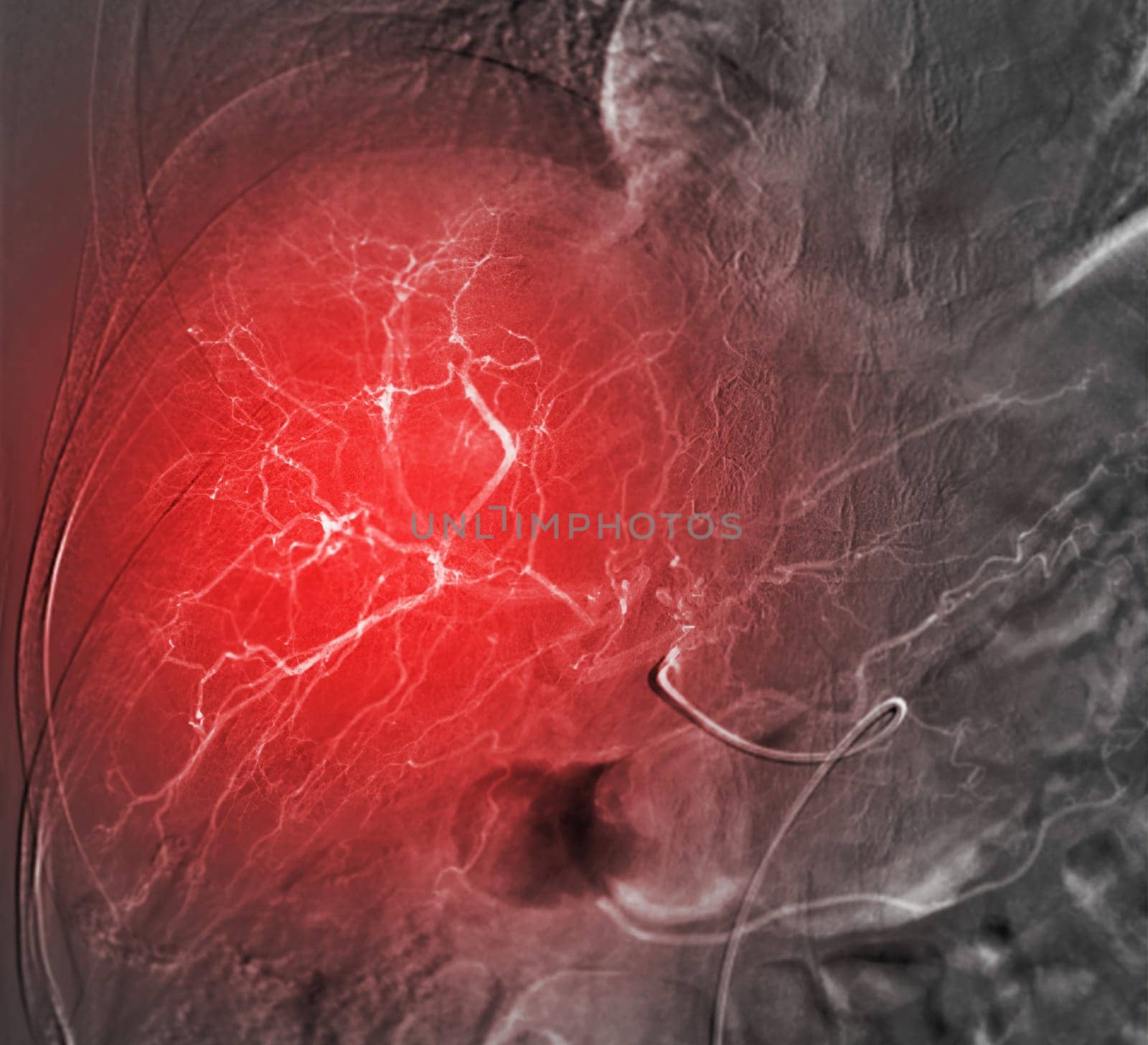
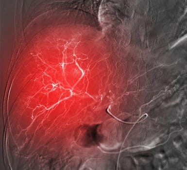
Imaging of TACE or Chemoembolization is a procedure that allows a dose of chemotherapy drugs to be administered directly to Liver tumor or HCC showing hepatic artery.
Stock PhotoUsername
samunellaResolution
3869x3520pxImaging of TACE or Chemoembolization is a procedure that allows a dose of chemotherapy drugs to be administered directly to Liver tumor or HCC showing hepatic artery.
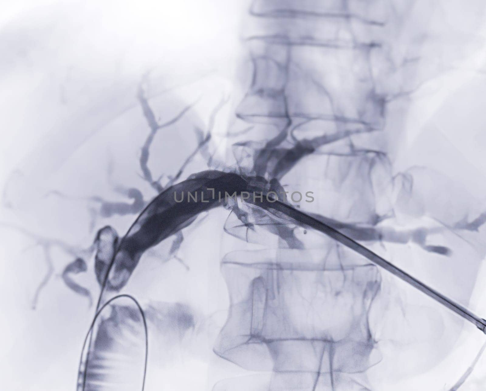
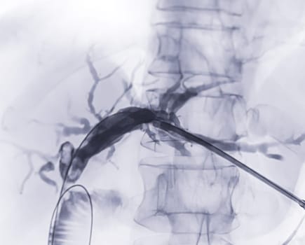
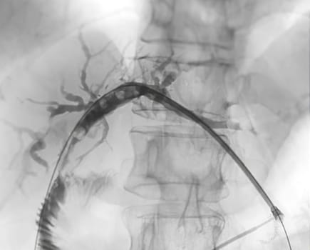
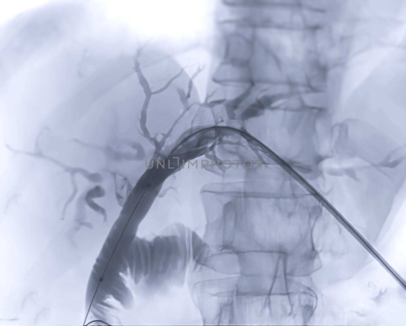
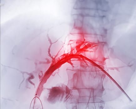
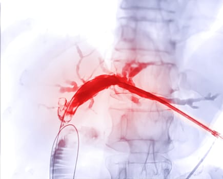
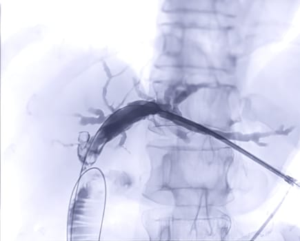
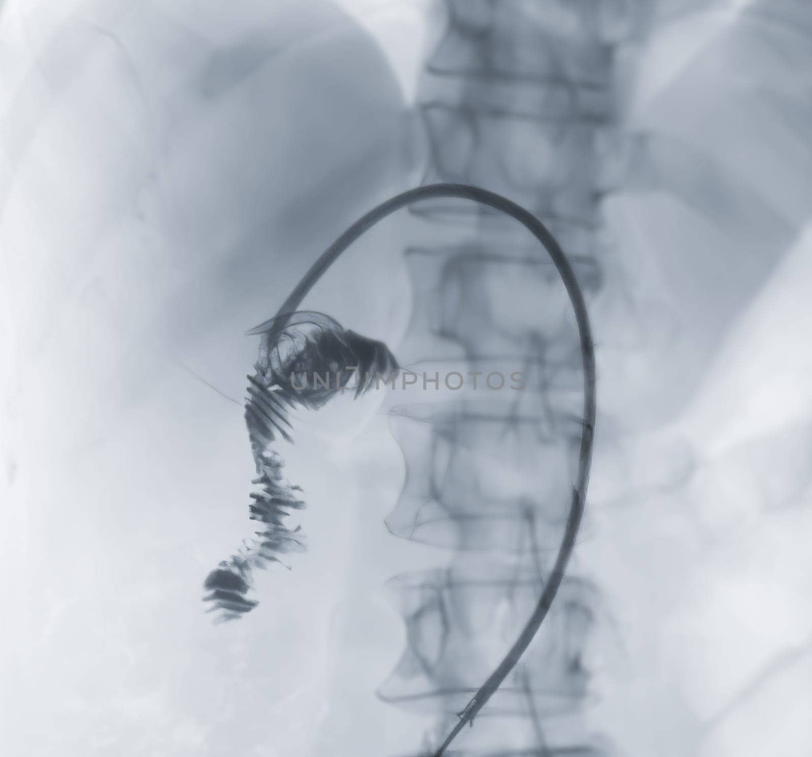
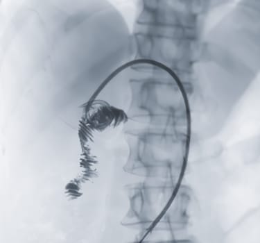
X-ray image of portal vein after Doctor doing ERCP and laparoscopic cholecystectomy inside modern operating room.
Stock PhotoUsername
samunellaResolution
3794x3052pxX-ray image of portal vein after Doctor doing ERCP and laparoscopic cholecystectomy inside modern operating room.

X-ray image of portal vein after Doctor doing ERCP and laparoscopic cholecystectomy inside modern operating room.
Stock PhotoUsername
samunellaResolution
3794x3052pxX-ray image of portal vein after Doctor doing ERCP and laparoscopic cholecystectomy inside modern operating room.
X-ray image of portal vein after Doctor doing ERCP and laparoscopic cholecystectomy inside modern operating room.
Stock PhotoUsername
samunellaResolution
3794x3052pxX-ray image of portal vein after Doctor doing ERCP and laparoscopic cholecystectomy inside modern operating room.


X-ray image of portal vein after Doctor doing ERCP and laparoscopic cholecystectomy inside modern operating room.
Stock PhotoUsername
samunellaResolution
3794x3052pxX-ray image of portal vein after Doctor doing ERCP and laparoscopic cholecystectomy inside modern operating room.

X-ray image of portal vein after Doctor doing ERCP and laparoscopic cholecystectomy inside modern operating room.
Stock PhotoUsername
samunellaResolution
3794x3052pxX-ray image of portal vein after Doctor doing ERCP and laparoscopic cholecystectomy inside modern operating room.
X-ray image of portal vein after Doctor doing ERCP and laparoscopic cholecystectomy inside modern operating room.
Stock PhotoUsername
samunellaResolution
3794x3052pxX-ray image of portal vein after Doctor doing ERCP and laparoscopic cholecystectomy inside modern operating room.

X-ray image of portal vein after Doctor doing ERCP and laparoscopic cholecystectomy inside modern operating room.
Stock PhotoUsername
samunellaResolution
3794x3052pxX-ray image of portal vein after Doctor doing ERCP and laparoscopic cholecystectomy inside modern operating room.
X-ray image of portal vein after Doctor doing ERCP and laparoscopic cholecystectomy inside modern operating room.
Stock PhotoUsername
samunellaResolution
3271x3052pxX-ray image of portal vein after Doctor doing ERCP and laparoscopic cholecystectomy inside modern operating room.
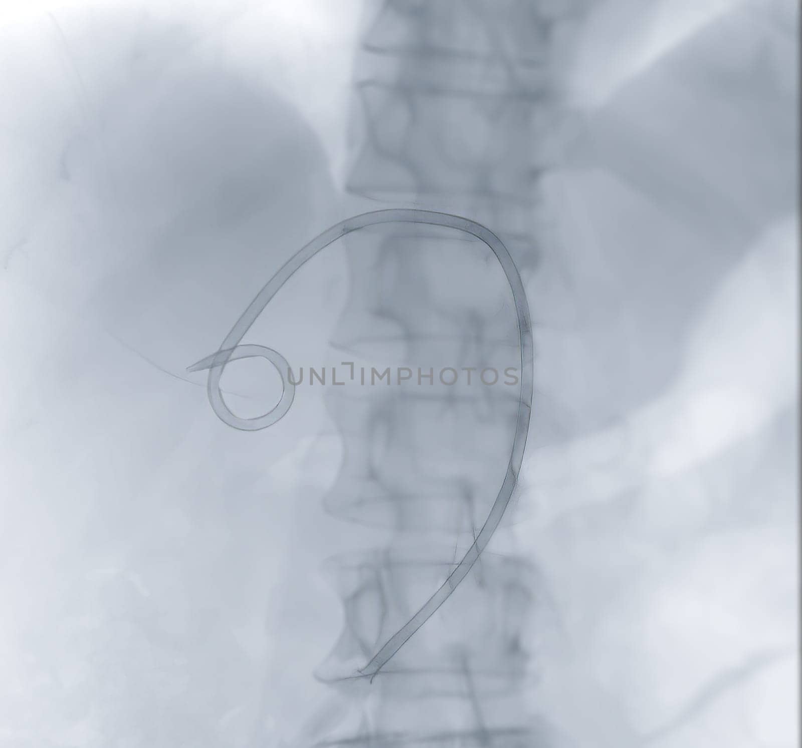
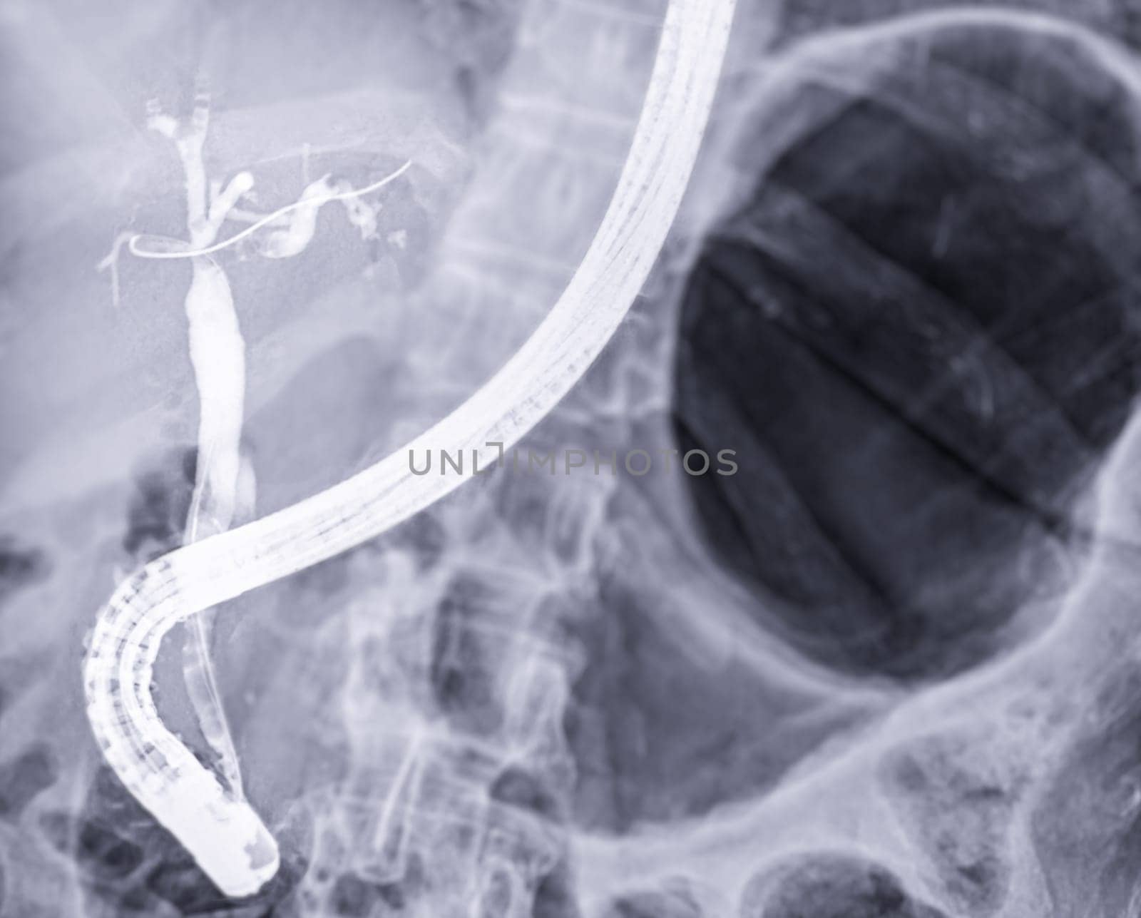
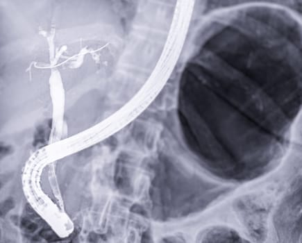
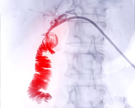
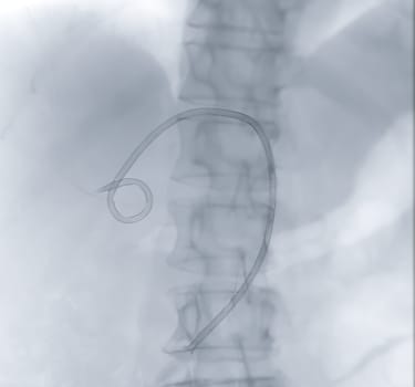
X-ray image of catheter in Upper abdomen.
Stock PhotoUsername
samunellaResolution
3271x3052pxX-ray image of catheter in Upper abdomen.

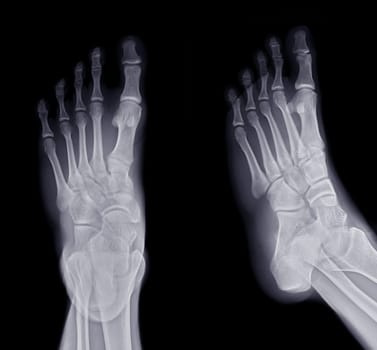
Foot x-ray image AP and Oblique view isolated on black background.
Stock PhotoUsername
samunellaResolution
5252x4882pxFoot x-ray image AP and Oblique view isolated on black background.
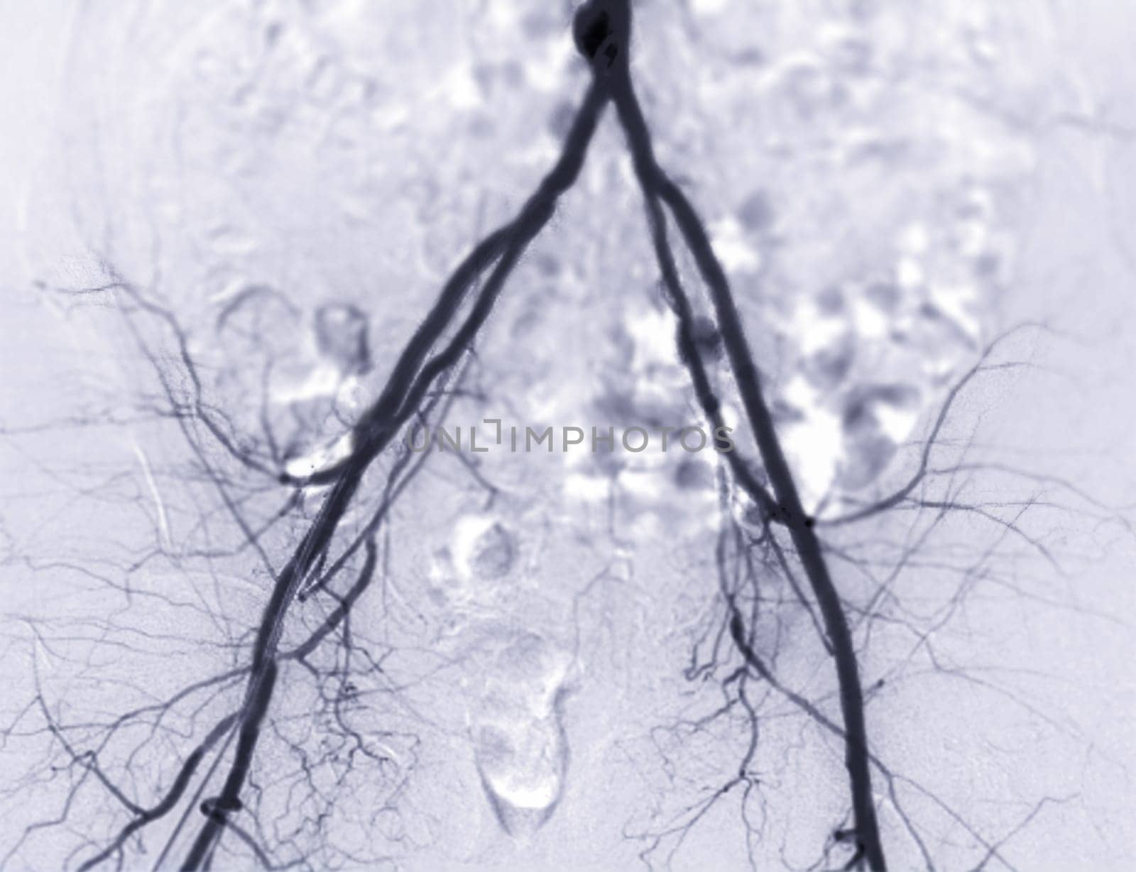
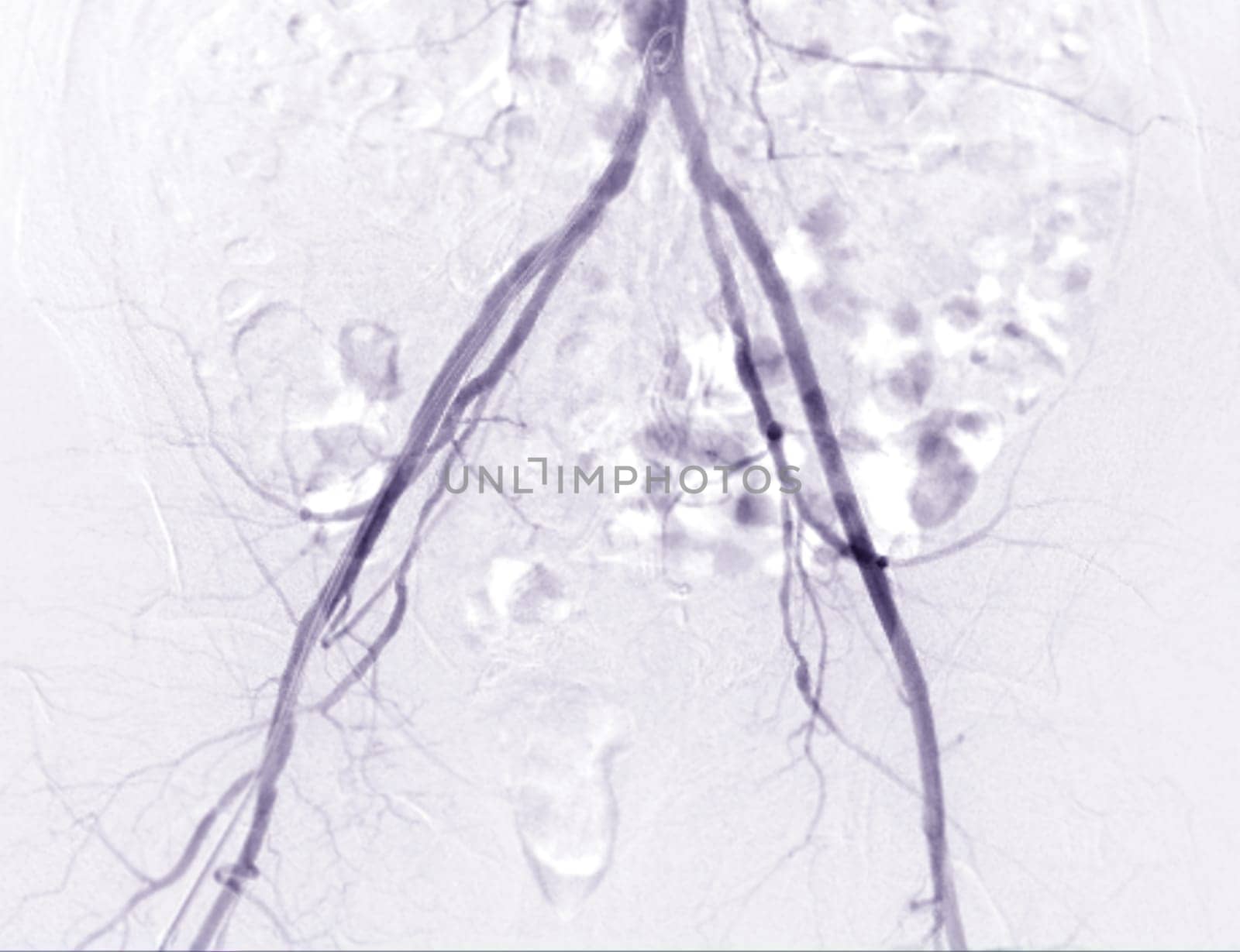
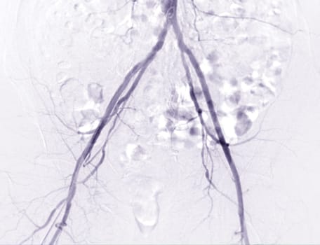
Femoral artery angiogram or angiography
Stock PhotoUsername
samunellaResolution
3984x3060pxFemoral artery angiogram or angiography
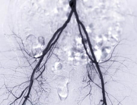
Femoral artery angiogram or angiography
Stock PhotoUsername
samunellaResolution
3984x3060pxFemoral artery angiogram or angiography
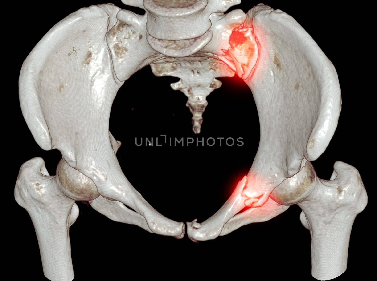
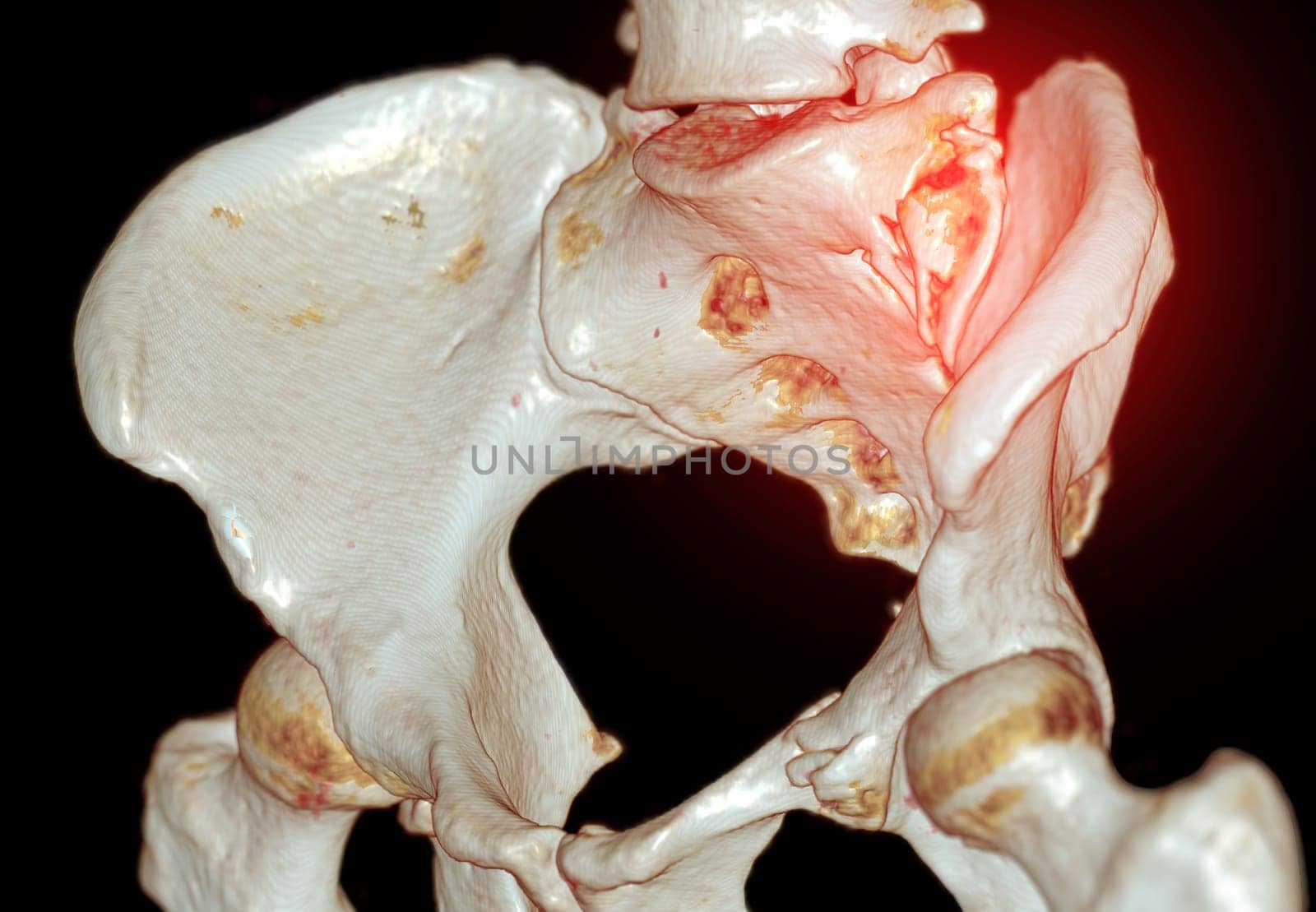
CT Scan pelvic bone with both hip joint 3D rendering showign fracture of sacrum and superior pubic rumus.
Stock PhotoUsername
samunellaResolution
4632x3456pxCT Scan pelvic bone with both hip joint 3D rendering showign fracture of sacrum and superior pubic rumus.
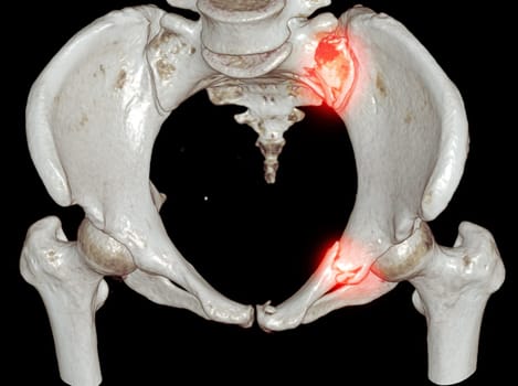
CT Scan pelvic bone with both hip joint 3D rendering showign fracture of sacrum and superior pubic rumus.
Stock PhotoUsername
samunellaResolution
3984x2761pxCT Scan pelvic bone with both hip joint 3D rendering showign fracture of sacrum and superior pubic rumus.

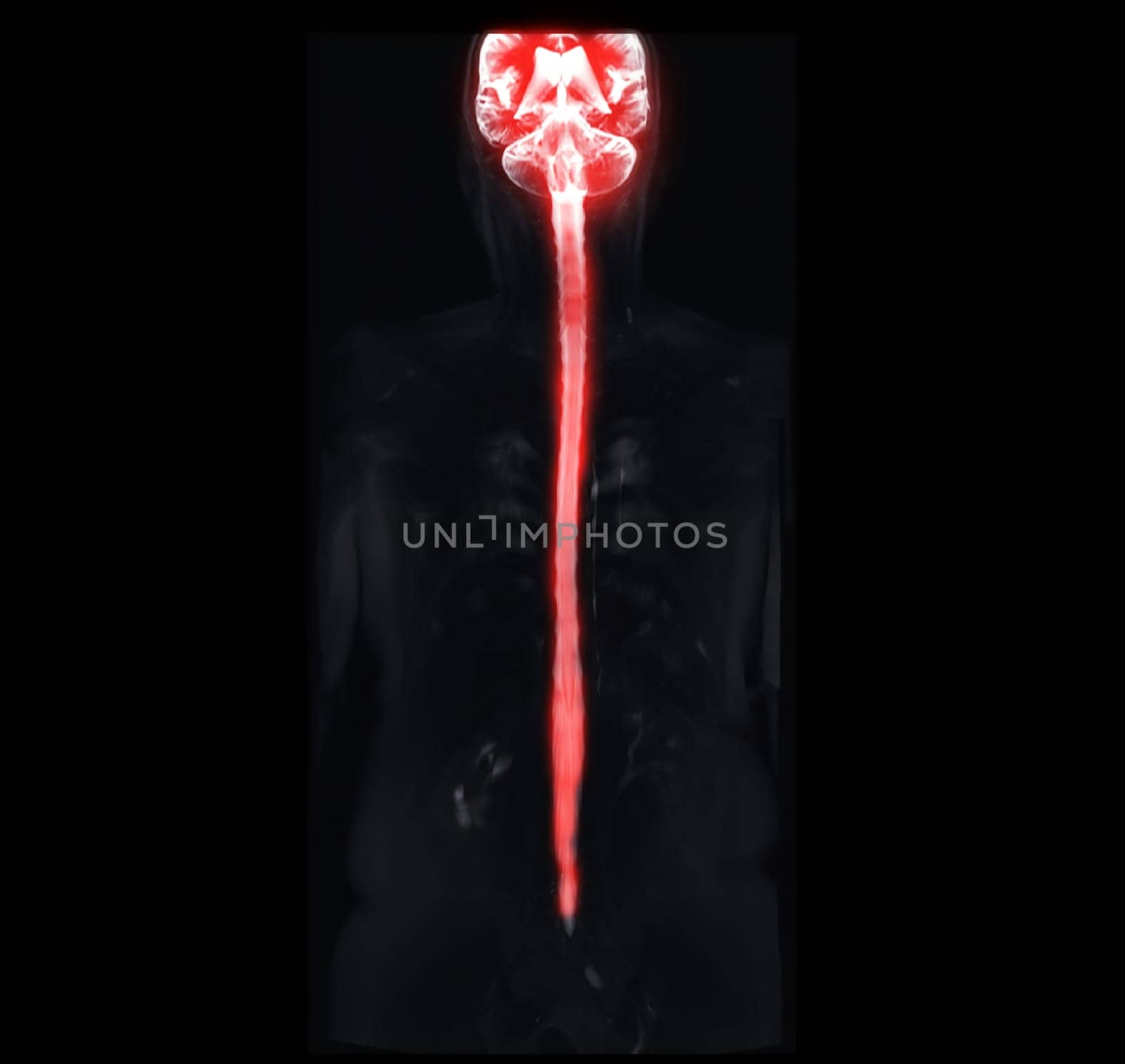
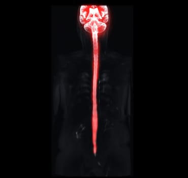
MRI of whole spine showing spinal cord.
Stock PhotoUsername
samunellaResolution
4056x3835pxMRI of whole spine showing spinal cord.
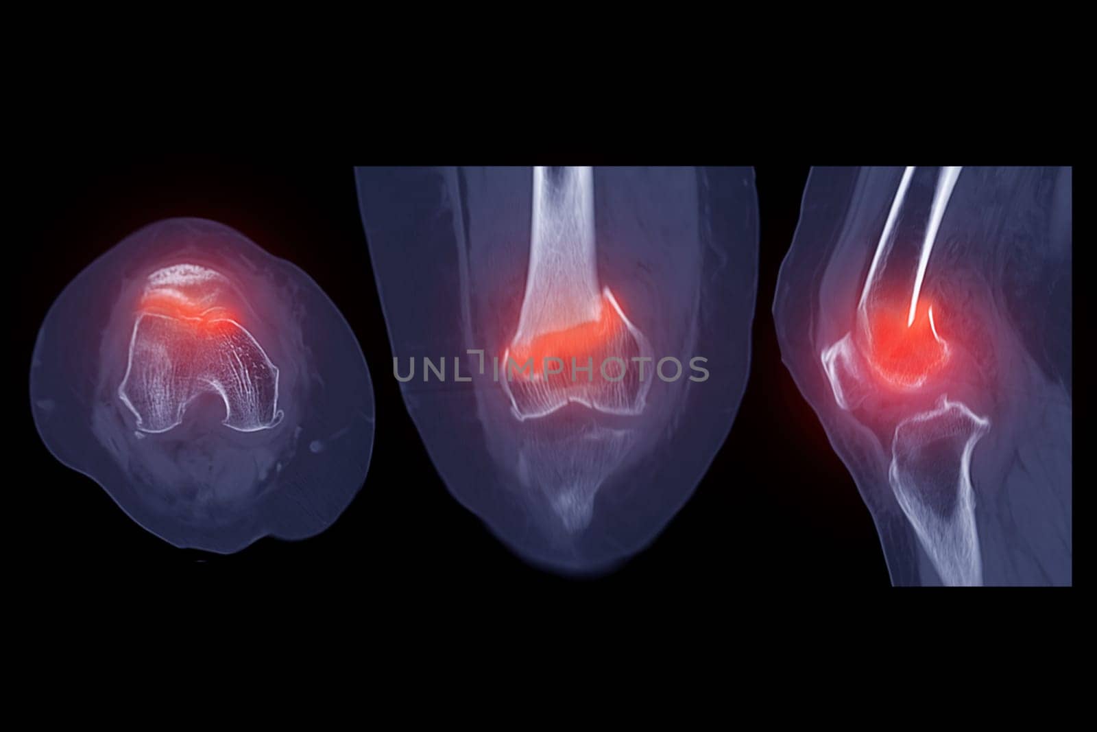
CT scan of knee joint 3D rendering image showing fracture of distal femur bone.
Stock PhotoUsername
samunellaResolution
6000x4000pxCT scan of knee joint 3D rendering image showing fracture of distal femur bone.

CT scan of knee joint 3D rendering image showing fracture of distal femur bone.
Stock PhotoUsername
samunellaResolution
6000x4000pxCT scan of knee joint 3D rendering image showing fracture of distal femur bone.
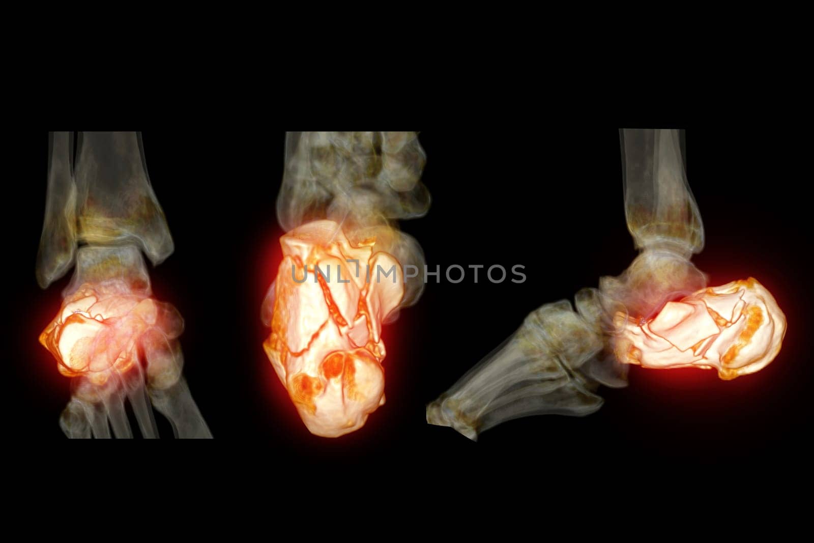
CT Scan ankle joint with 3d rendering of calcaneus bone showing Calcaneus (Heel Bone) Fractures.
Stock PhotoUsername
samunellaResolution
6000x4000pxCT Scan ankle joint with 3d rendering of calcaneus bone showing Calcaneus (Heel Bone) Fractures.

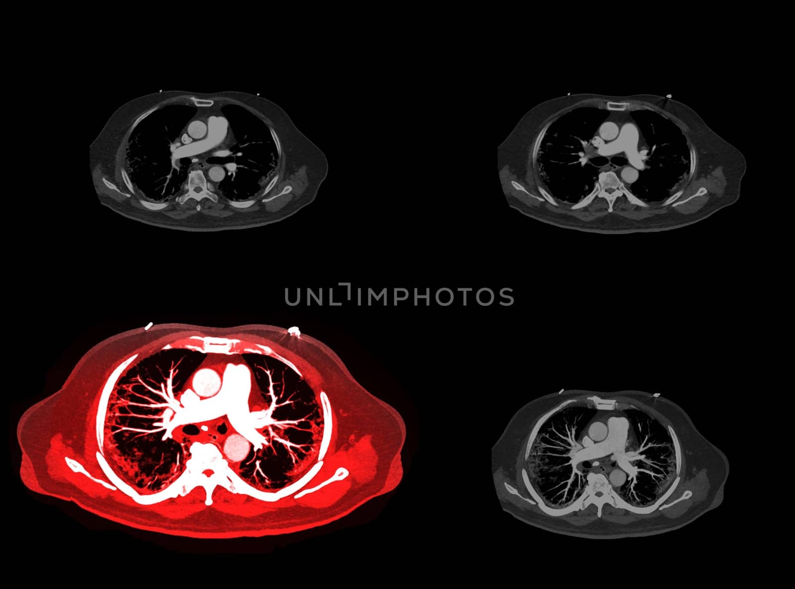
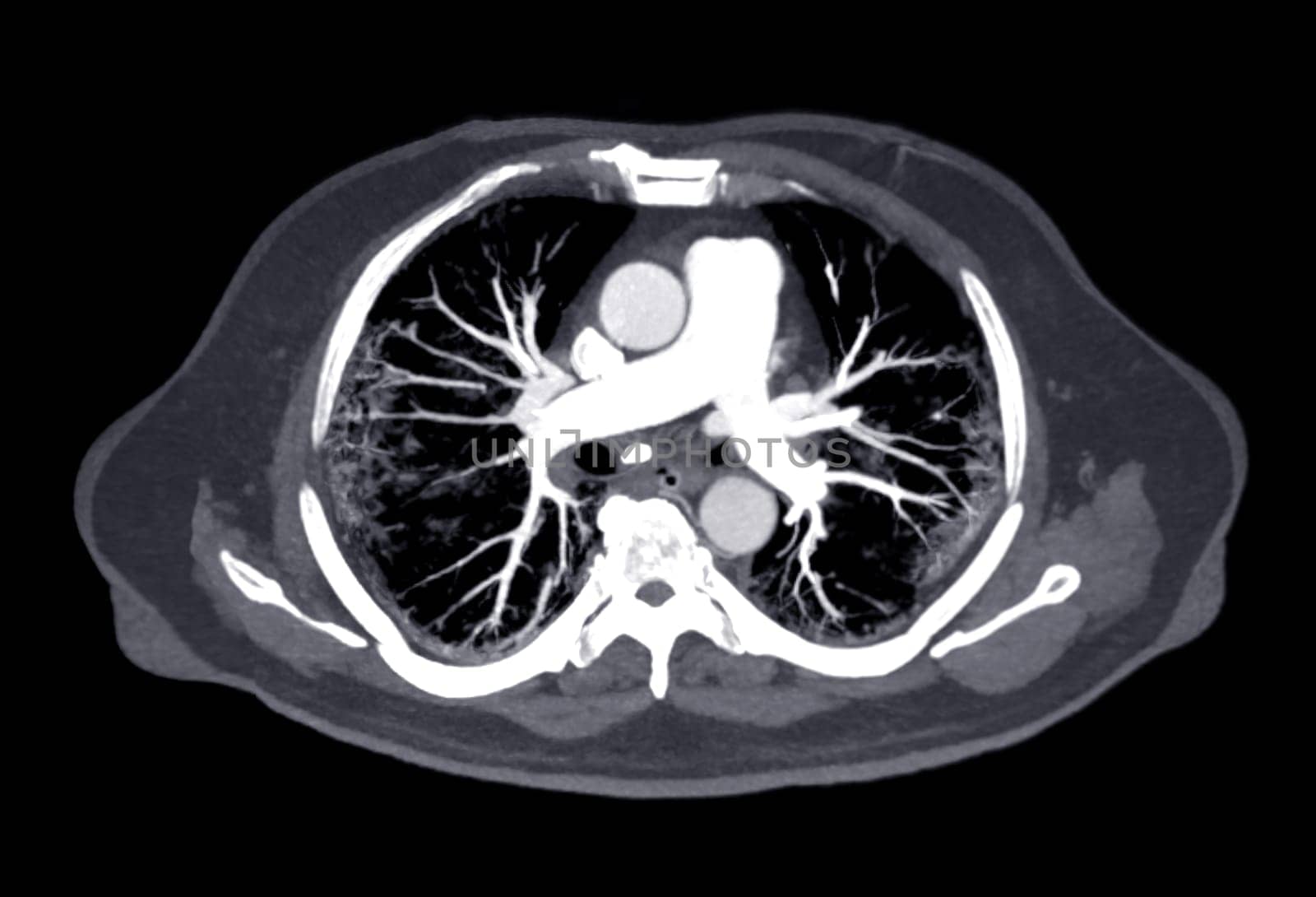
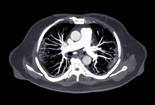
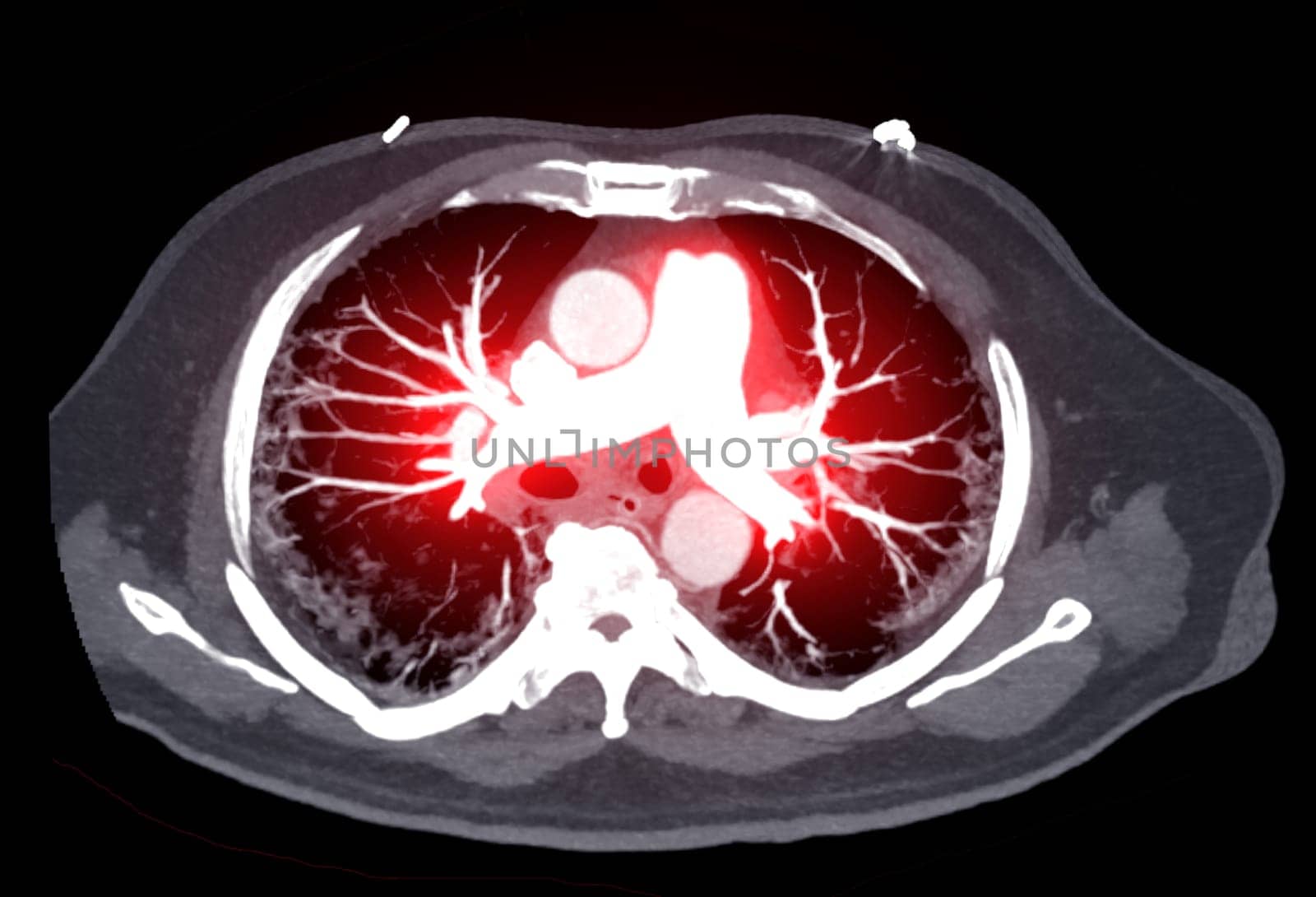
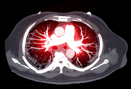
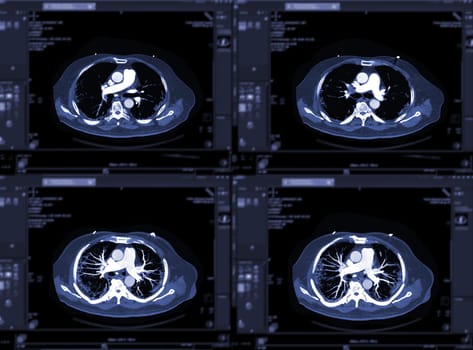
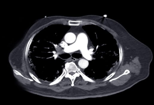
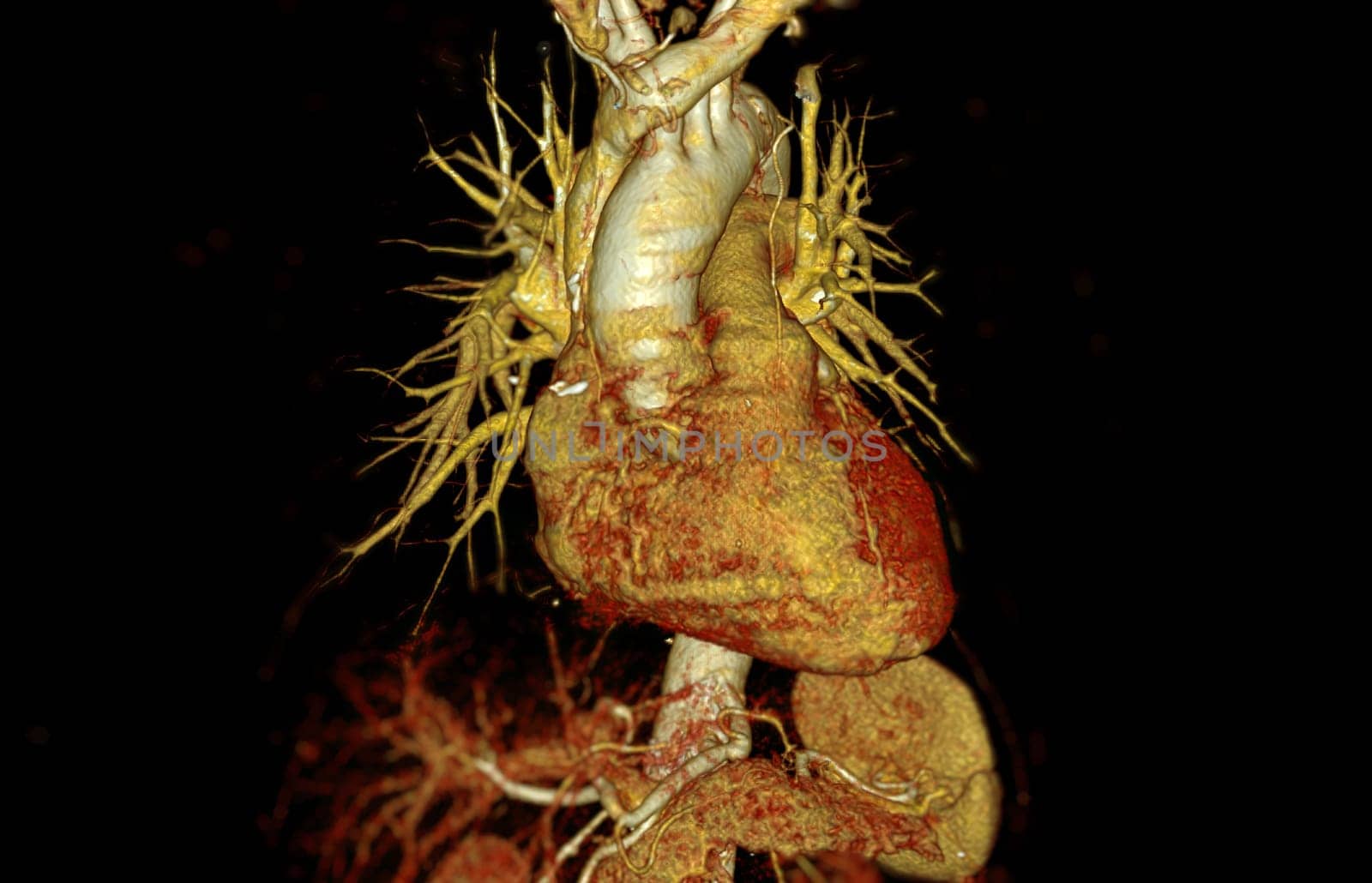
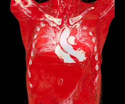
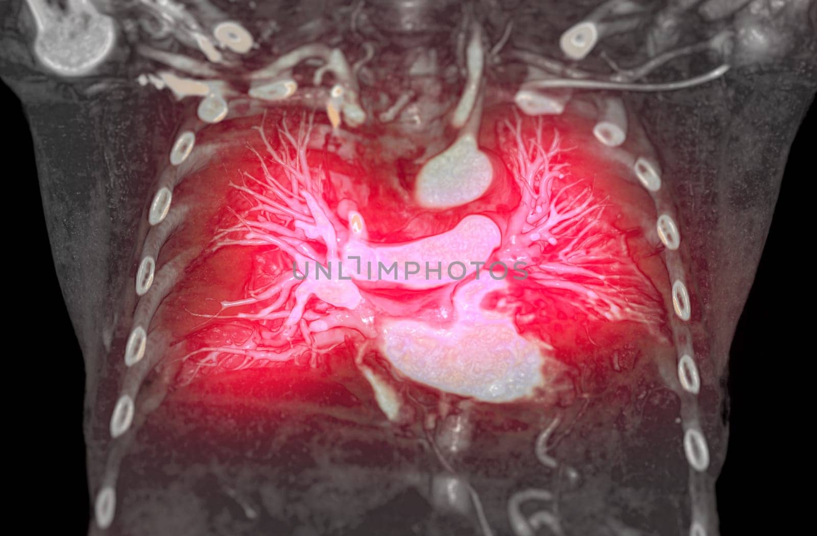
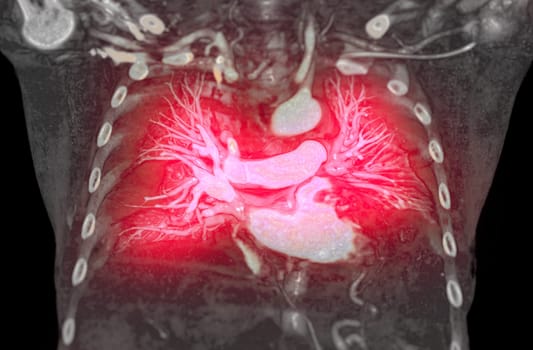
CTA pulmonary arteries 3D rendering showing branch of pulmonary artery
Stock PhotoUsername
samunellaResolution
5888x4363pxCTA pulmonary arteries 3D rendering showing branch of pulmonary artery
CTA pulmonary arteries 3D rendering showing branch of pulmonary artery
Stock PhotoUsername
samunellaResolution
5206x3548pxCTA pulmonary arteries 3D rendering showing branch of pulmonary artery
CTA pulmonary arteries 3D rendering showing branch of pulmonary artery
Stock PhotoUsername
samunellaResolution
5206x3548pxCTA pulmonary arteries 3D rendering showing branch of pulmonary artery

CTA pulmonary arteries 3D rendering showing branch of pulmonary artery
Stock PhotoUsername
samunellaResolution
5888x4363pxCTA pulmonary arteries 3D rendering showing branch of pulmonary artery

CTA pulmonary arteries 3D rendering showing branch of pulmonary artery
Stock PhotoUsername
samunellaResolution
5206x3548pxCTA pulmonary arteries 3D rendering showing branch of pulmonary artery

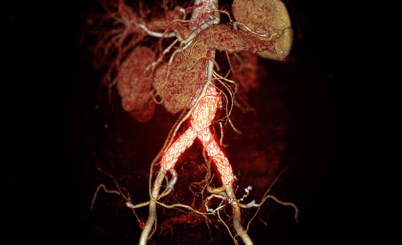
CTA of the aorta with stent-grafting in patient Abdominal aortic aneurysm.
Stock PhotoUsername
samunellaResolution
3748x2292pxCTA of the aorta with stent-grafting in patient Abdominal aortic aneurysm.
CTA of the aorta with stent-grafting in patient Abdominal aortic aneurysm.
Stock PhotoUsername
samunellaResolution
3748x2412pxCTA of the aorta with stent-grafting in patient Abdominal aortic aneurysm.


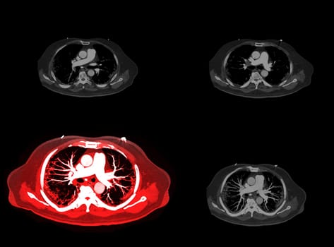
CTA pulmonary arteries 3D rendering showing branch of pulmonary artery
Stock PhotoUsername
samunellaResolution
4038x3347pxCTA pulmonary arteries 3D rendering showing branch of pulmonary artery
CTA pulmonary arteries 3D rendering showing branch of pulmonary artery
Stock PhotoUsername
samunellaResolution
4707x3088pxCTA pulmonary arteries 3D rendering showing branch of pulmonary artery

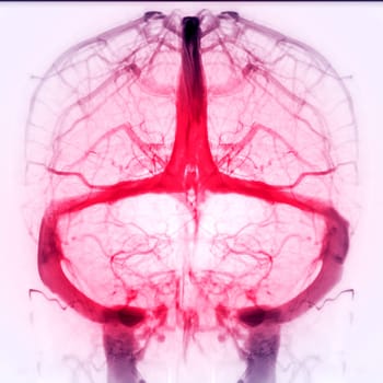
Cerebral Venograpgy for diagnosisi Cerebral Venous Thrombosis
Stock PhotoUsername
samunellaResolution
3898x3892pxCerebral Venograpgy for diagnosisi Cerebral Venous Thrombosis