- Filter By:
-
-
Stock photos and images of username:samunella

X-ray of Elbow showing internal fixation of the elbow joint.
Stock PhotoUsername
samunellaResolution
4462x4818pxX-ray of Elbow showing internal fixation of the elbow joint.
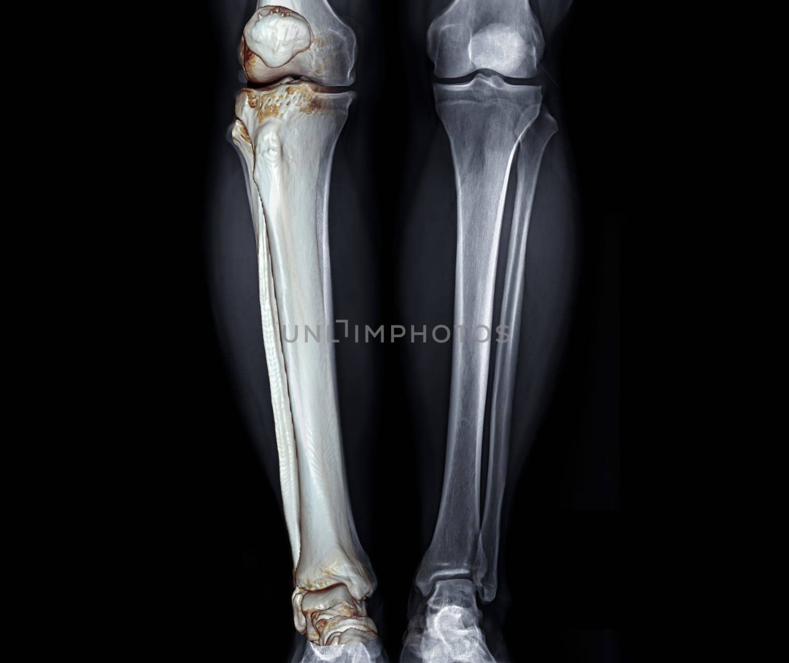
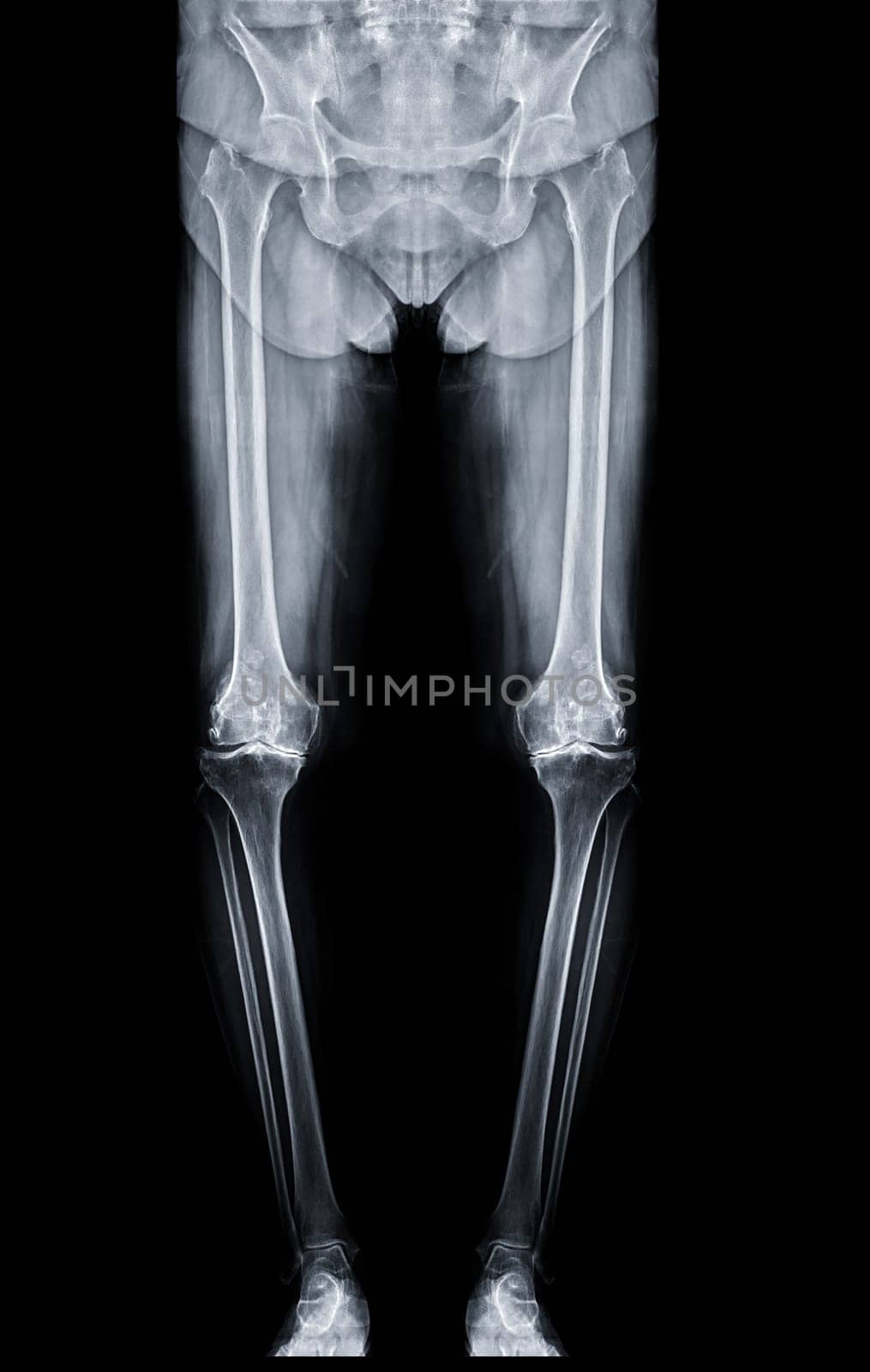
X-ray image of both Leg AP view with 3D leg for diagnostic knee fracrure.
Stock PhotoUsername
samunellaResolution
3832x3222pxX-ray image of both Leg AP view with 3D leg for diagnostic knee fracrure.

Happy Valentines Day with 3d rendering of red heart balloons shapes with rose and light bulbs on red background. Gift card, love party, invitation voucher design, poster template.
Stock PhotoUsername
samunellaResolution
4000x3000pxHappy Valentines Day with 3d rendering of red heart balloons shapes with rose and light bulbs on red background. Gift card, love party, invitation voucher design, poster template.

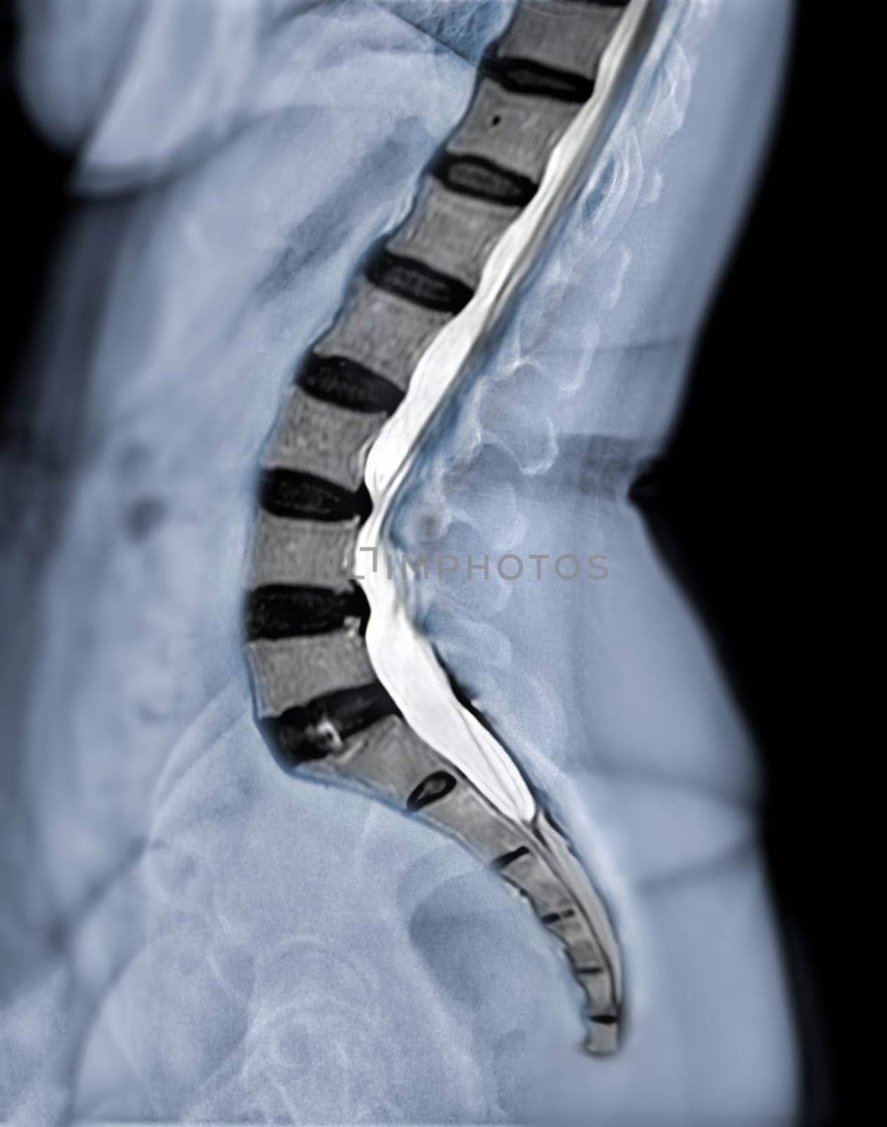
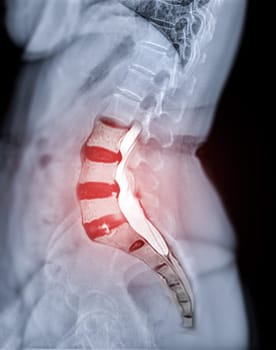
X-ray image of lumbar Spine or L-s spine lateral view with MRI l-s spine for diagnosis lower back pain.
Stock PhotoUsername
samunellaResolution
5242x6658pxX-ray image of lumbar Spine or L-s spine lateral view with MRI l-s spine for diagnosis lower back pain.
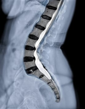

X-ray image of lumbar Spine or L-s spine lateral view with MRI l-s spine for diagnosis lower back pain.
Stock PhotoUsername
samunellaResolution
5242x6658pxX-ray image of lumbar Spine or L-s spine lateral view with MRI l-s spine for diagnosis lower back pain.
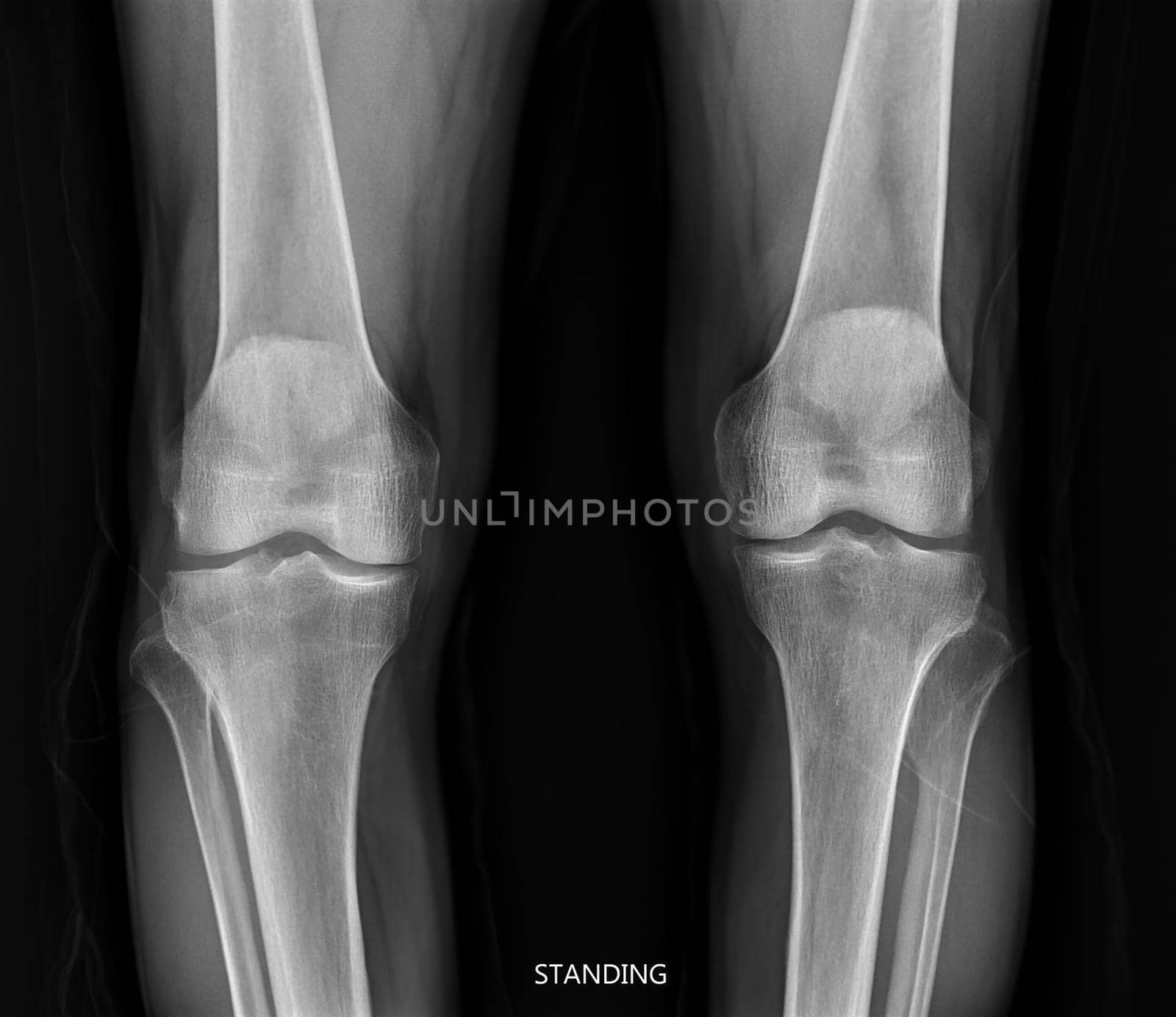
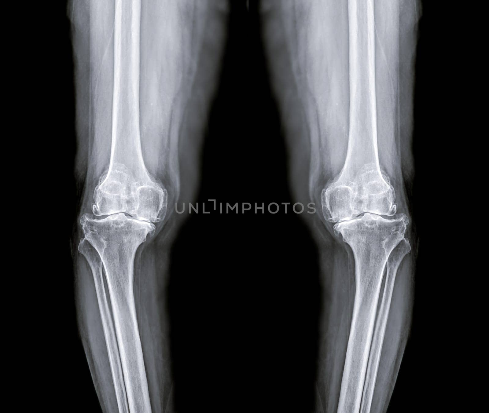
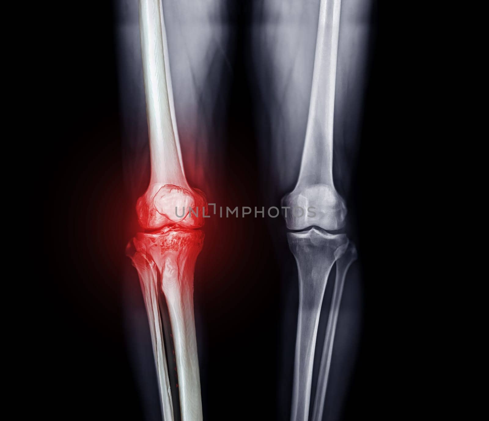
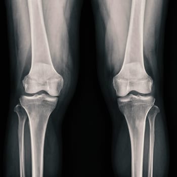
film x-ray both knee joint AP view for diagnosis knee pain from osteoarthritis knee and fracture .
Stock PhotoUsername
samunellaResolution
6474x5600pxfilm x-ray both knee joint AP view for diagnosis knee pain from osteoarthritis knee and fracture .

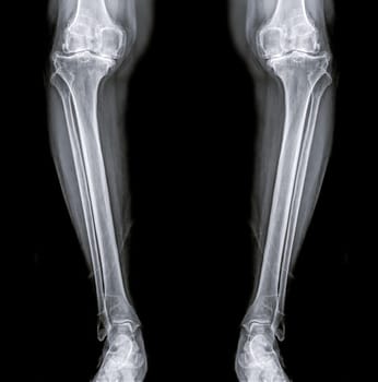
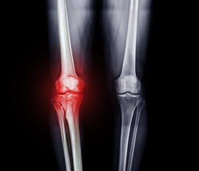
film x-ray both knee joint AP view for diagnosis knee pain from osteoarthritis knee and fracture .
Stock PhotoUsername
samunellaResolution
3474x2936pxfilm x-ray both knee joint AP view for diagnosis knee pain from osteoarthritis knee and fracture .
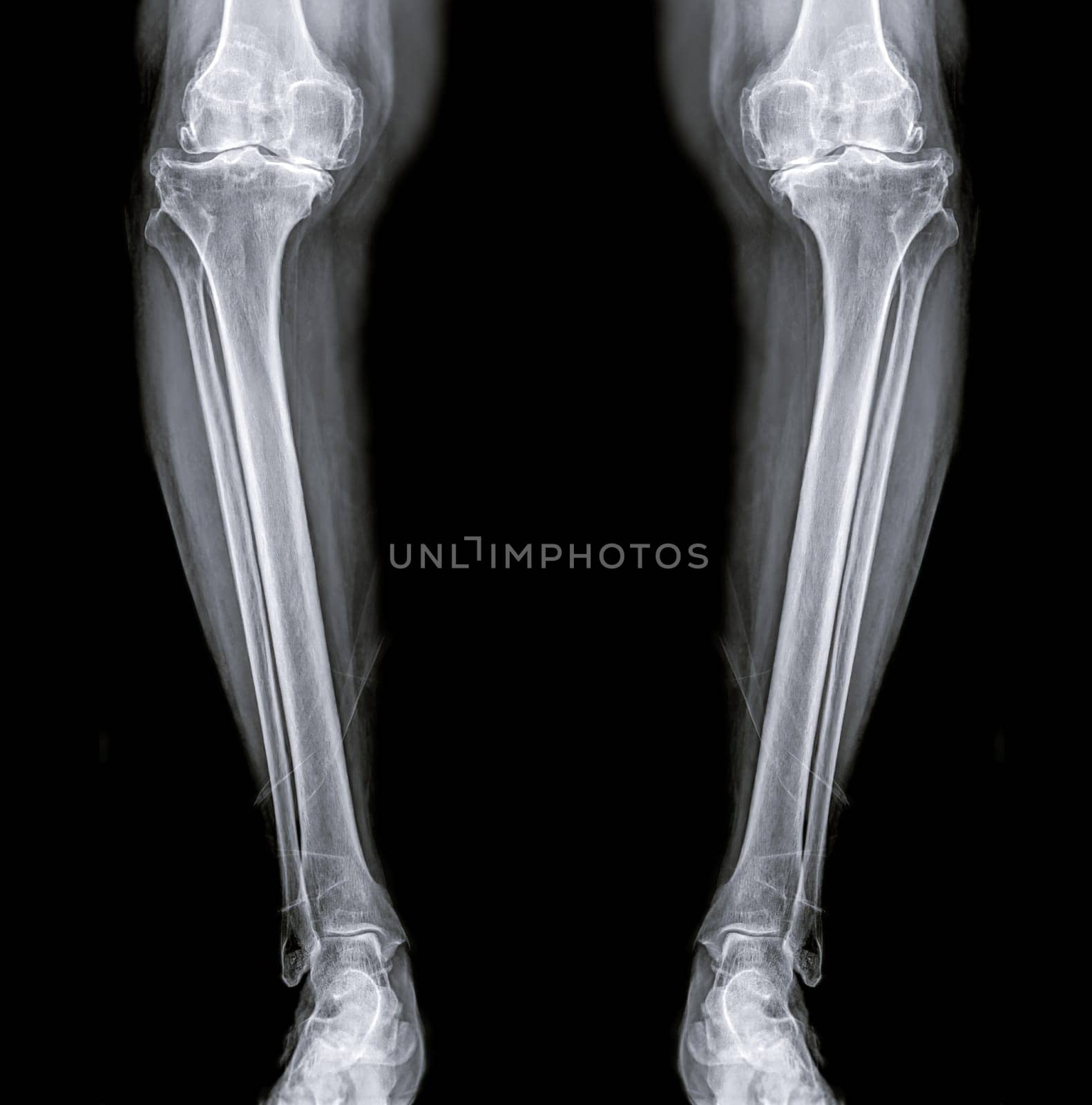
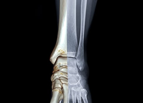
film x-ray both Leg AP view for diagnosis knee pain from osteoarthritis knee and fracture .
Stock PhotoUsername
samunellaResolution
3170x3208pxfilm x-ray both Leg AP view for diagnosis knee pain from osteoarthritis knee and fracture .
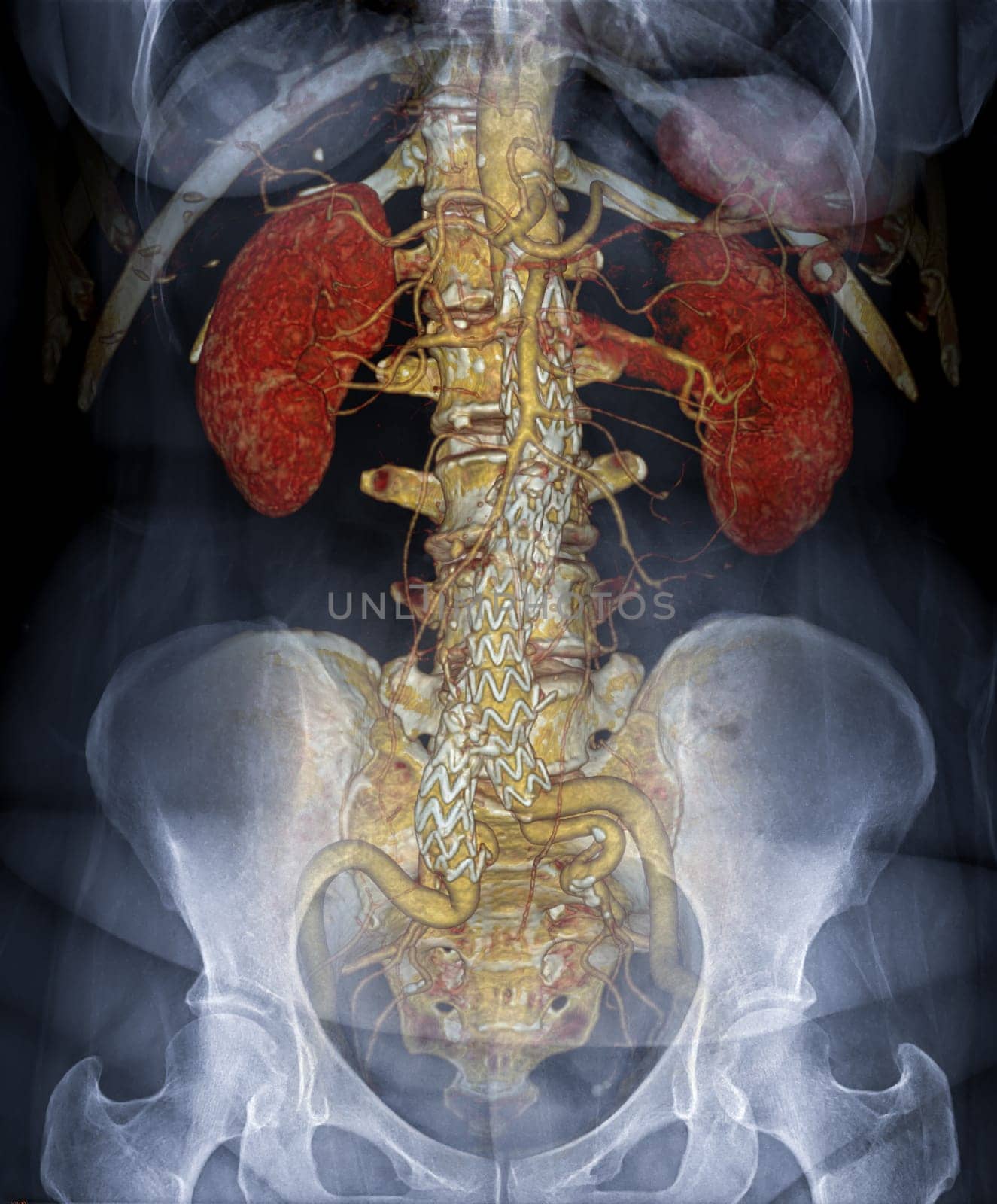
CTA ABDOMINAL AORTA 3D rendering fusion with X-ray Abdomen image.
Stock PhotoUsername
samunellaResolution
5540x6688pxCTA ABDOMINAL AORTA 3D rendering fusion with X-ray Abdomen image.


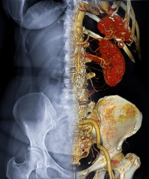
CTA ABDOMINAL AORTA 3D rendering fusion with X-ray Abdomen image.
Stock PhotoUsername
samunellaResolution
5540x6688pxCTA ABDOMINAL AORTA 3D rendering fusion with X-ray Abdomen image.

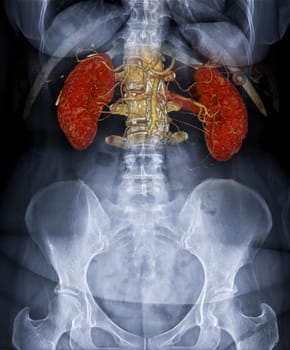
CTA ABDOMINAL AORTA 3D rendering fusion with X-ray Abdomen image.
Stock PhotoUsername
samunellaResolution
5540x6688pxCTA ABDOMINAL AORTA 3D rendering fusion with X-ray Abdomen image.
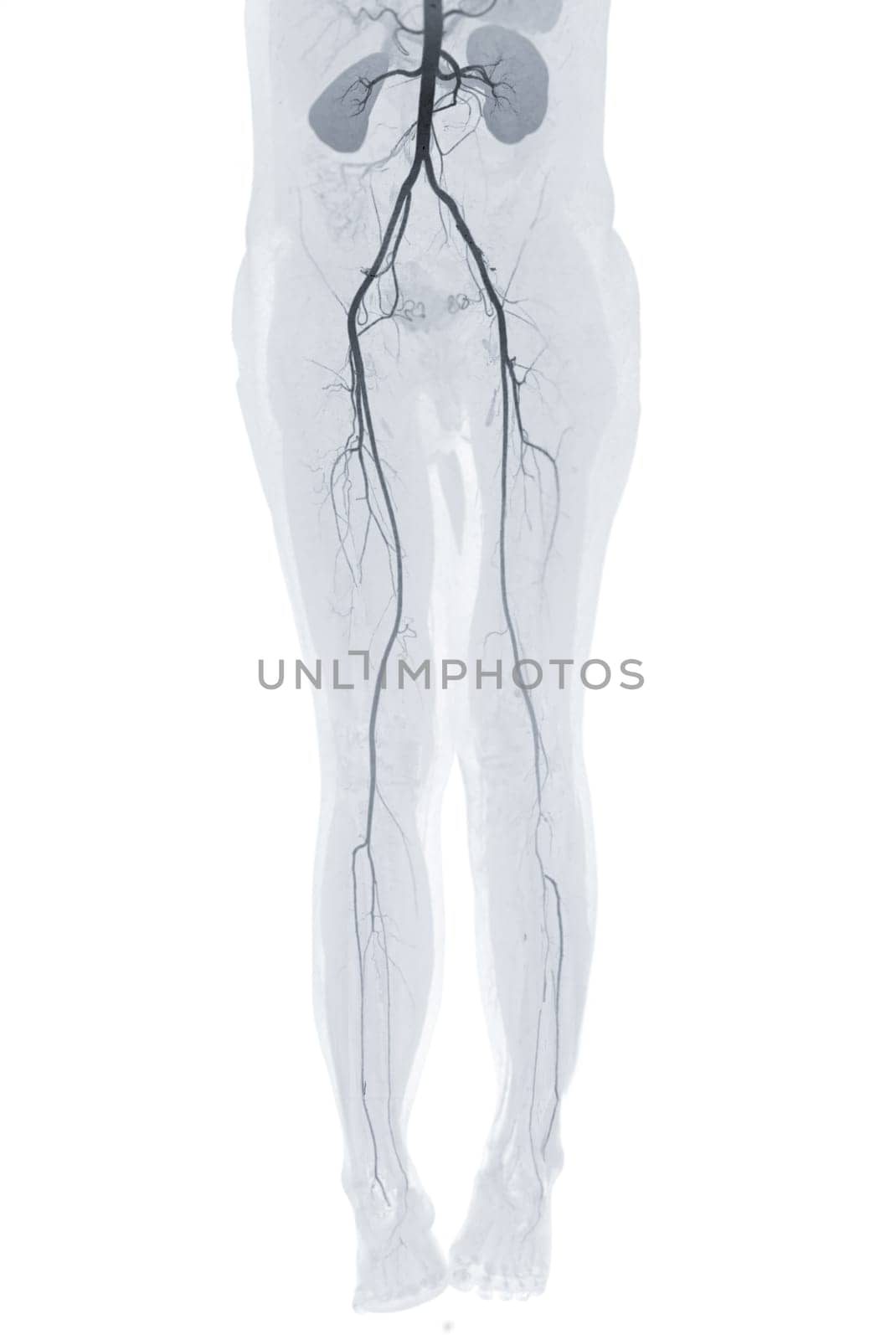
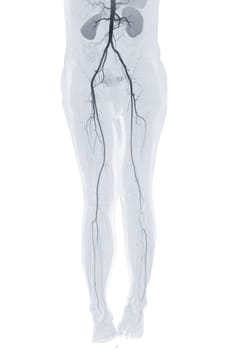
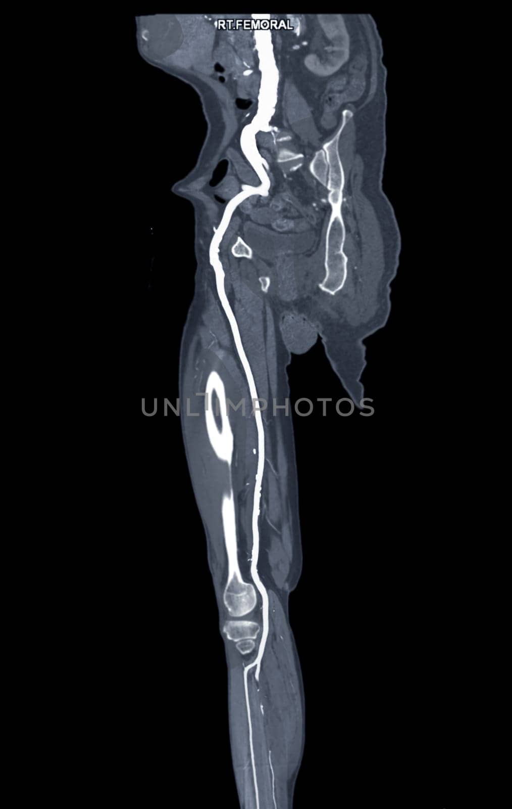
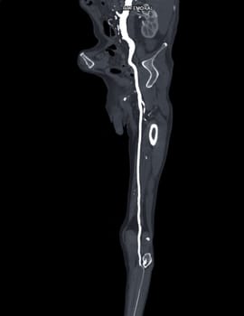
CTA femoral artery run off showing femoral artery Presenting with Acute or Chronic Peripheral Arterial Disease.
Stock PhotoUsername
samunellaResolution
3878x5800pxCTA femoral artery run off showing femoral artery Presenting with Acute or Chronic Peripheral Arterial Disease.

x-ray Both hip ap view showing Right hip replacement or hip prosthesis made from titanium with fusion 3D rendering .
Stock PhotoUsername
samunellaResolution
8146x5604pxx-ray Both hip ap view showing Right hip replacement or hip prosthesis made from titanium with fusion 3D rendering .

Love Rose gold Foil Balloon 3D rendering isolated on white background. Clipping path.
Stock PhotoUsername
samunellaResolution
4000x3000pxLove Rose gold Foil Balloon 3D rendering isolated on white background. Clipping path.


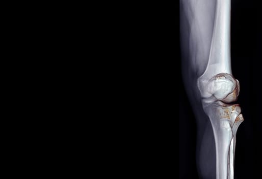
X-ray of knee joint AP view for diagnosis knee pain from osteoarthritis knee and fracture .
Stock PhotoUsername
samunellaResolution
3452x5124pxX-ray of knee joint AP view for diagnosis knee pain from osteoarthritis knee and fracture .
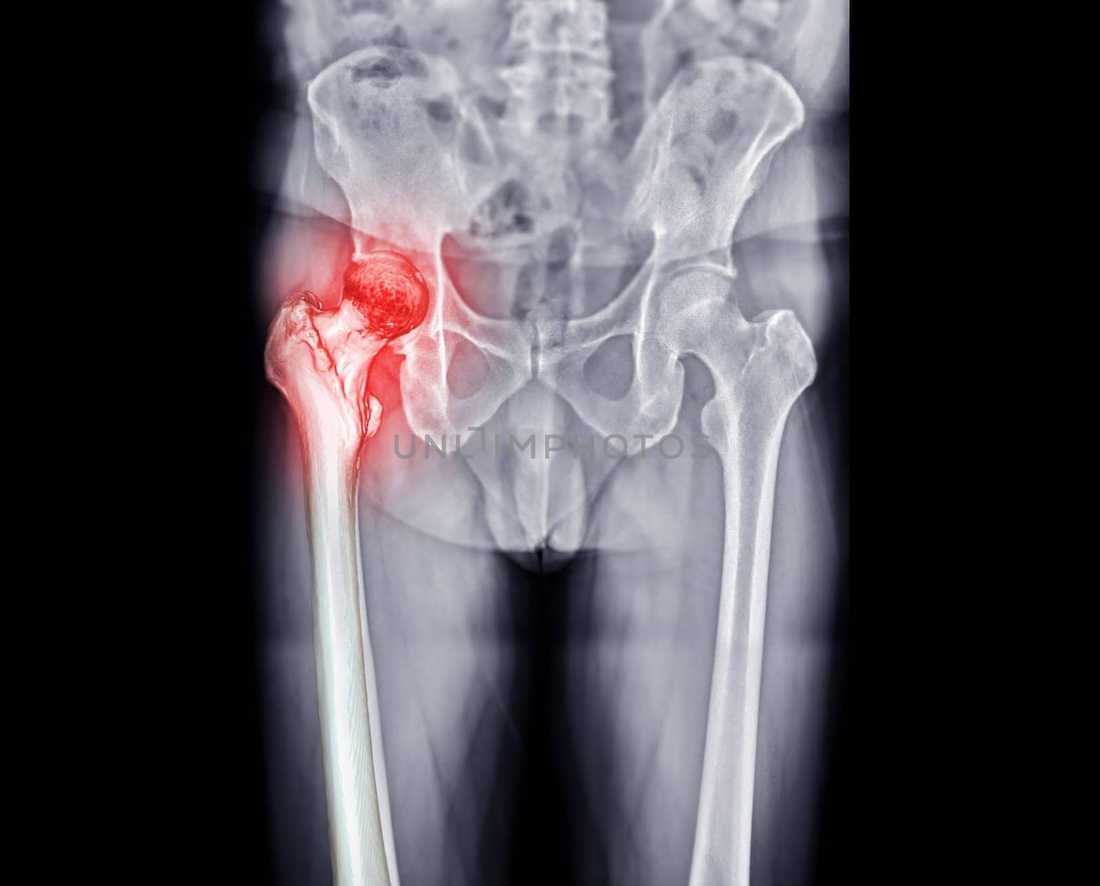
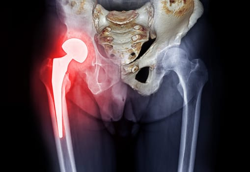
x-ray Both hip ap view showing Right hip replacement or hip prosthesis made from titanium .
Stock PhotoUsername
samunellaResolution
4136x3330pxx-ray Both hip ap view showing Right hip replacement or hip prosthesis made from titanium .

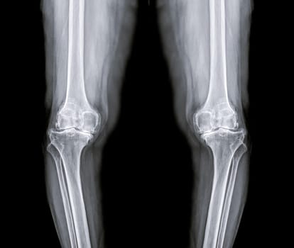
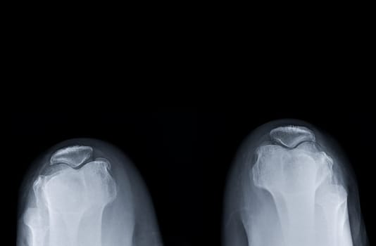
X-ray image of both knee AP view for diagnostic Osteoarthritis or knee fracrure.
Stock PhotoUsername
samunellaResolution
4136x3563pxX-ray image of both knee AP view for diagnostic Osteoarthritis or knee fracrure.

X-ray of knee joint fusion with 3D rendering .
Stock PhotoUsername
samunellaResolution
5692x4101pxX-ray of knee joint fusion with 3D rendering .
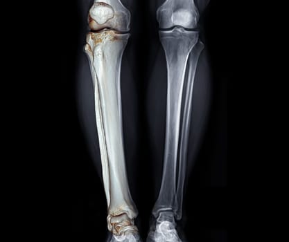
X-ray of knee joint and CT knee 3D rendering .
Stock PhotoUsername
samunellaResolution
5692x4101pxX-ray of knee joint and CT knee 3D rendering .

X-ray of knee joint fusion with 3D rendering .
Stock PhotoUsername
samunellaResolution
3452x5124pxX-ray of knee joint fusion with 3D rendering .
X-ray image of Left wrist joint AP and Lateral view for showing fracture of radius bone.
Stock PhotoUsername
samunellaResolution
2544x3198pxX-ray image of Left wrist joint AP and Lateral view for showing fracture of radius bone.
X-ray image of Left wrist joint AP and Lateral view for showing fracture of radius bone.
Stock PhotoUsername
samunellaResolution
2544x3198pxX-ray image of Left wrist joint AP and Lateral view for showing fracture of radius bone.

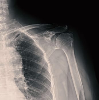
X-ray Shoulder joint shoulder transcapular view for diagnosis fracture of shoulder joint.
Stock PhotoUsername
samunellaResolution
3714x4282pxX-ray Shoulder joint shoulder transcapular view for diagnosis fracture of shoulder joint.
X-ray Shoulder joint shoulder transcapular view for diagnosis fracture of shoulder joint.
Stock PhotoUsername
samunellaResolution
4626x4666pxX-ray Shoulder joint shoulder transcapular view for diagnosis fracture of shoulder joint.
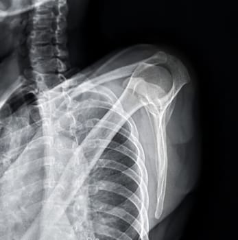
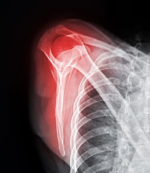
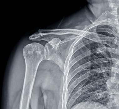
X-ray Shoulder joint shoulder front view for diagnosis fracture of shoulder joint.
Stock PhotoUsername
samunellaResolution
3230x3274pxX-ray Shoulder joint shoulder front view for diagnosis fracture of shoulder joint.

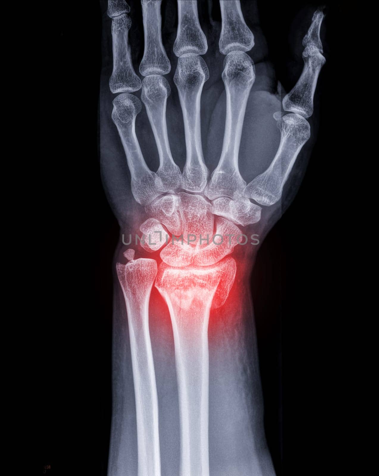
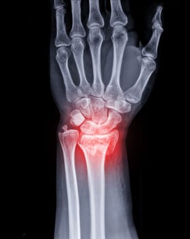
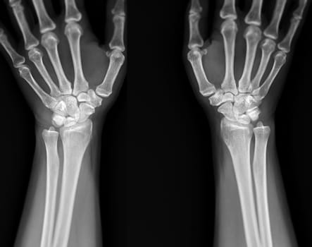
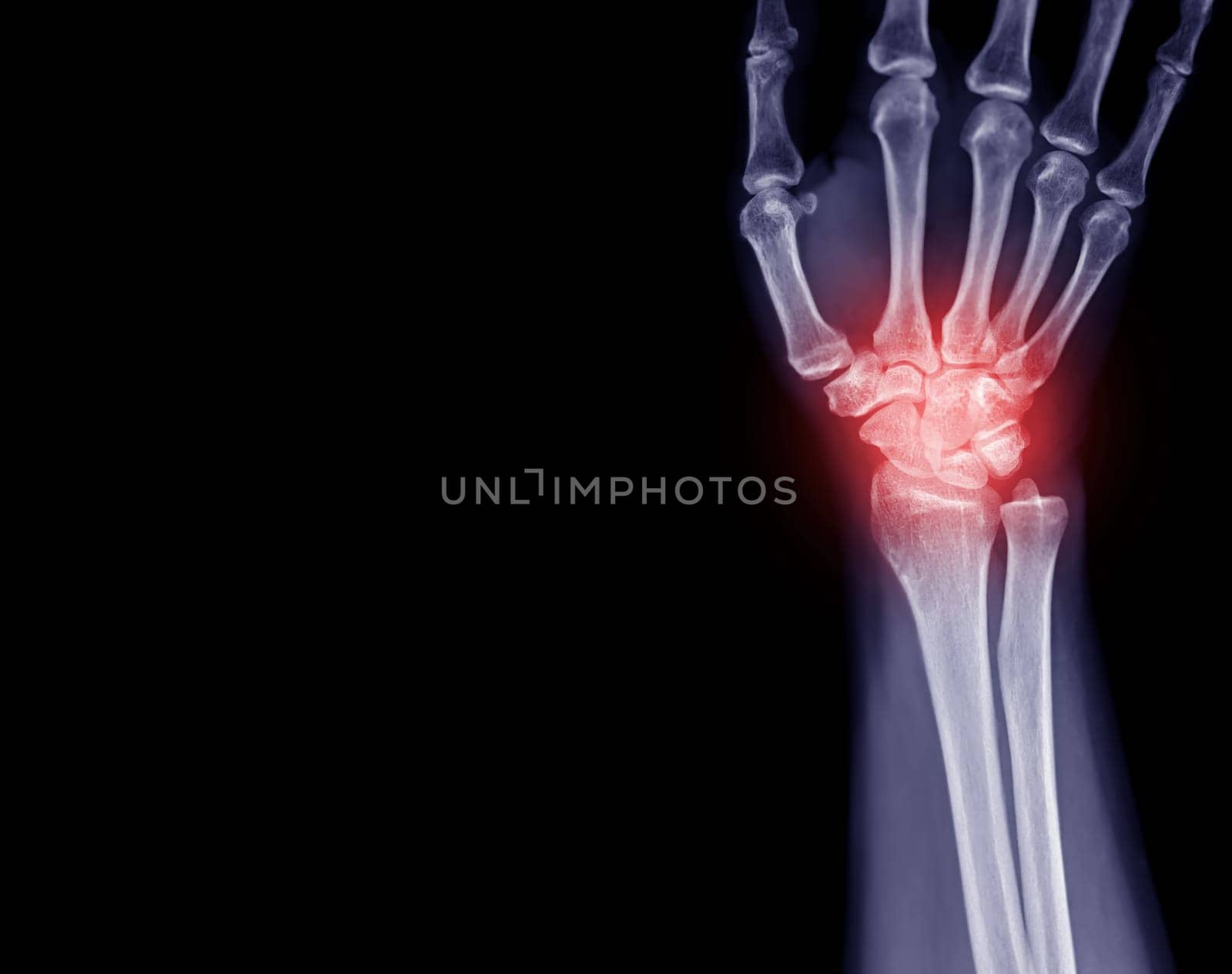
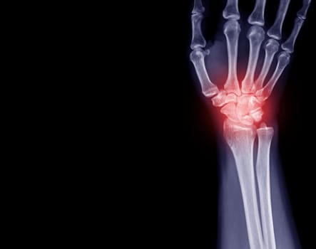
X-ray image of wrist joint front view of normal wrist joint.
Stock PhotoUsername
samunellaResolution
4294x3396pxX-ray image of wrist joint front view of normal wrist joint.
X-ray image of wrist joint front view of normal wrist joint.
Stock PhotoUsername
samunellaResolution
4294x3396pxX-ray image of wrist joint front view of normal wrist joint.
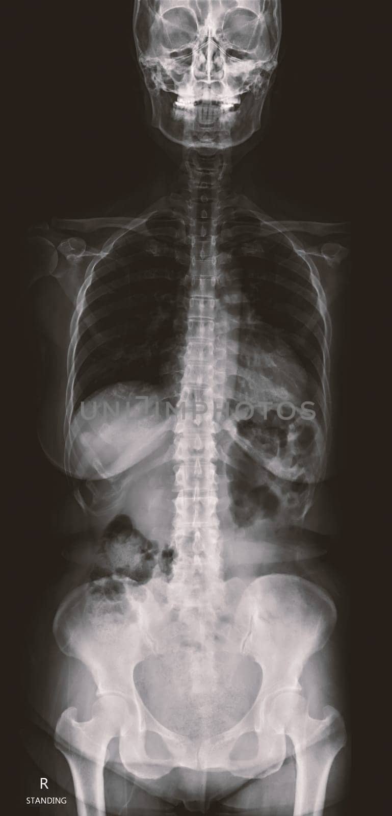
X-ray image of Whole Spine for diagnosis scoliosis of spine.
Stock PhotoUsername
samunellaResolution
2642x5496pxX-ray image of Whole Spine for diagnosis scoliosis of spine.
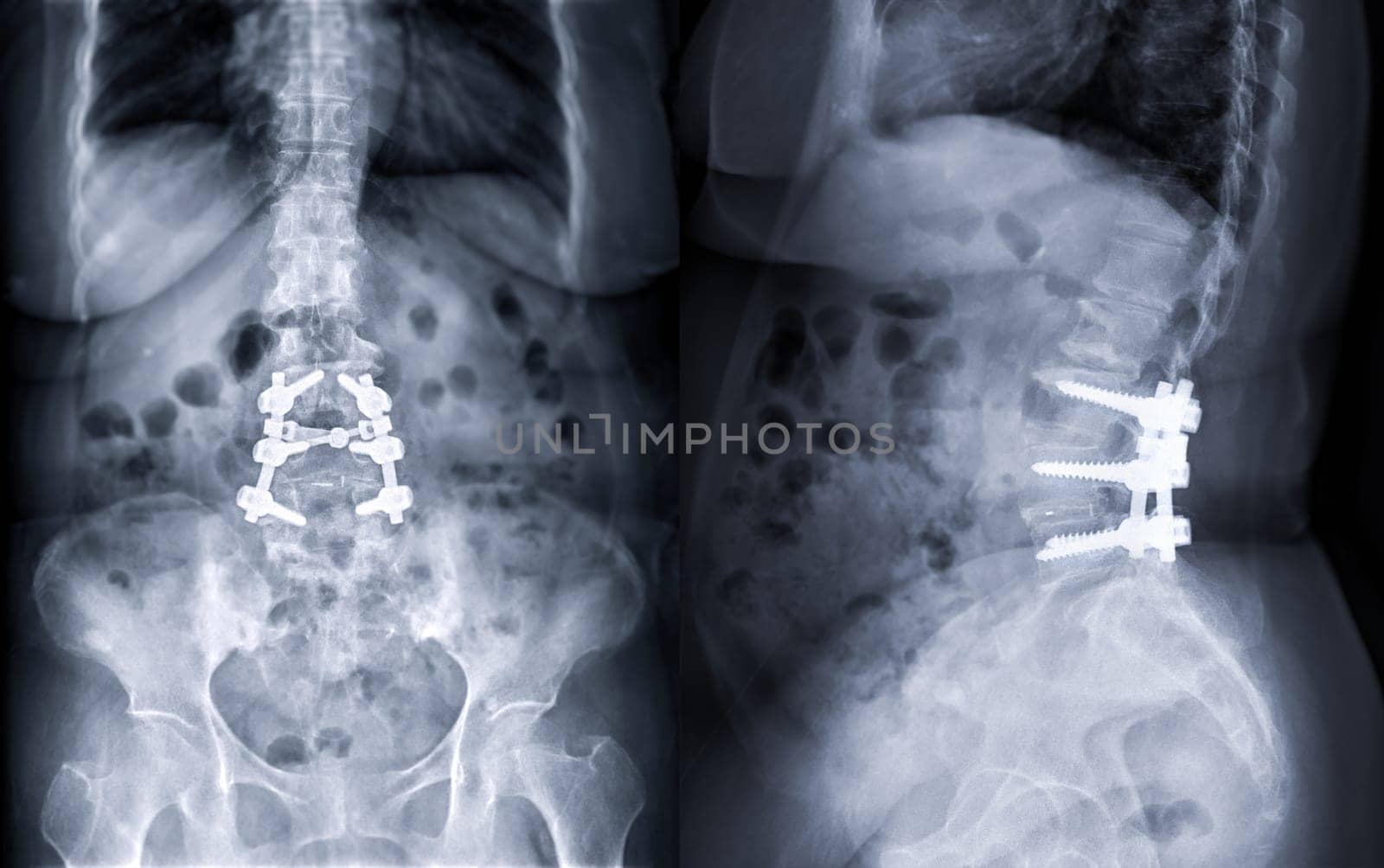
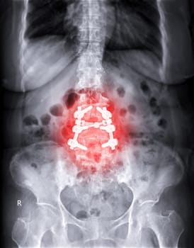
X-ray image of Lumbar spine showing pedicle screw fixation and decompression surgery in patient with spinal canal stenosis.
Stock PhotoUsername
samunellaResolution
6240x3914pxX-ray image of Lumbar spine showing pedicle screw fixation and decompression surgery in patient with spinal canal stenosis.

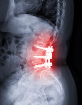
X-ray image of Lumbar spine showing pedicle screw fixation and decompression surgery in patient with spinal canal stenosis.
Stock PhotoUsername
samunellaResolution
5334x6816pxX-ray image of Lumbar spine showing pedicle screw fixation and decompression surgery in patient with spinal canal stenosis.

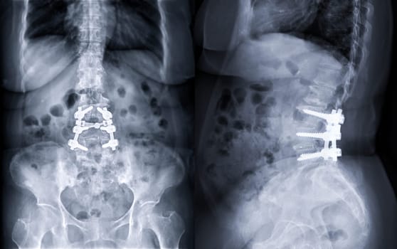
X-ray image of Lumbar spine showing pedicle screw fixation and decompression surgery in patient with spinal canal stenosis.
Stock PhotoUsername
samunellaResolution
5334x6816pxX-ray image of Lumbar spine showing pedicle screw fixation and decompression surgery in patient with spinal canal stenosis.
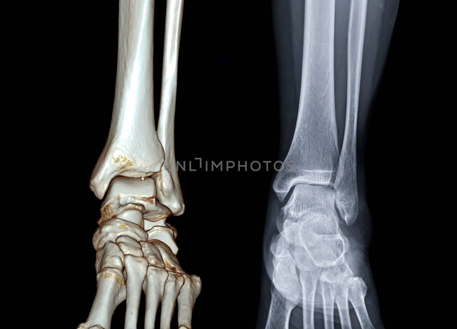
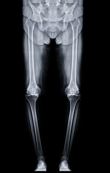
Scanogram is a Full-length standing AP radiograph of both lower extremities including the hip, knee, and ankle.
Stock PhotoUsername
samunellaResolution
4017x6328pxScanogram is a Full-length standing AP radiograph of both lower extremities including the hip, knee, and ankle.

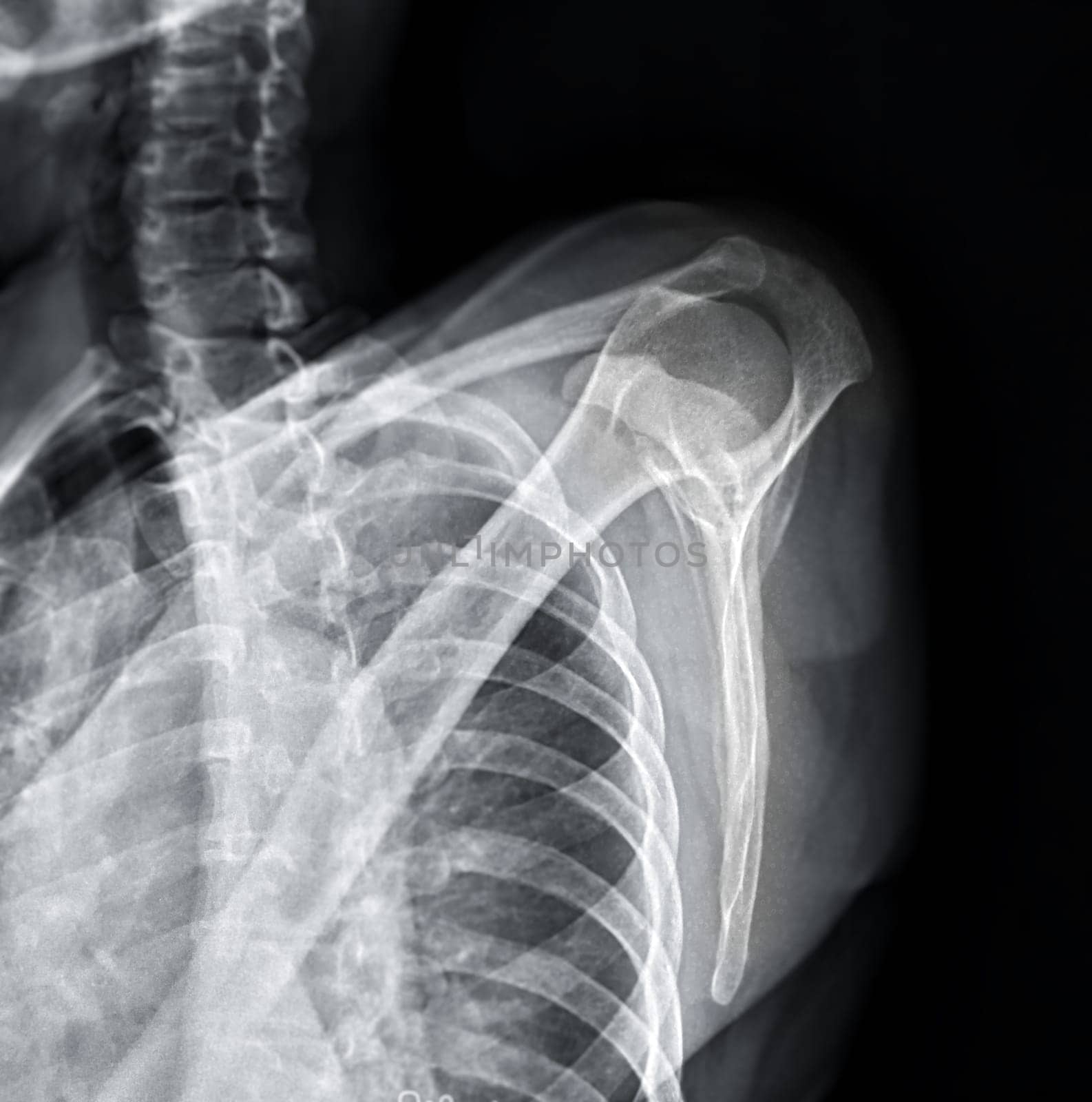
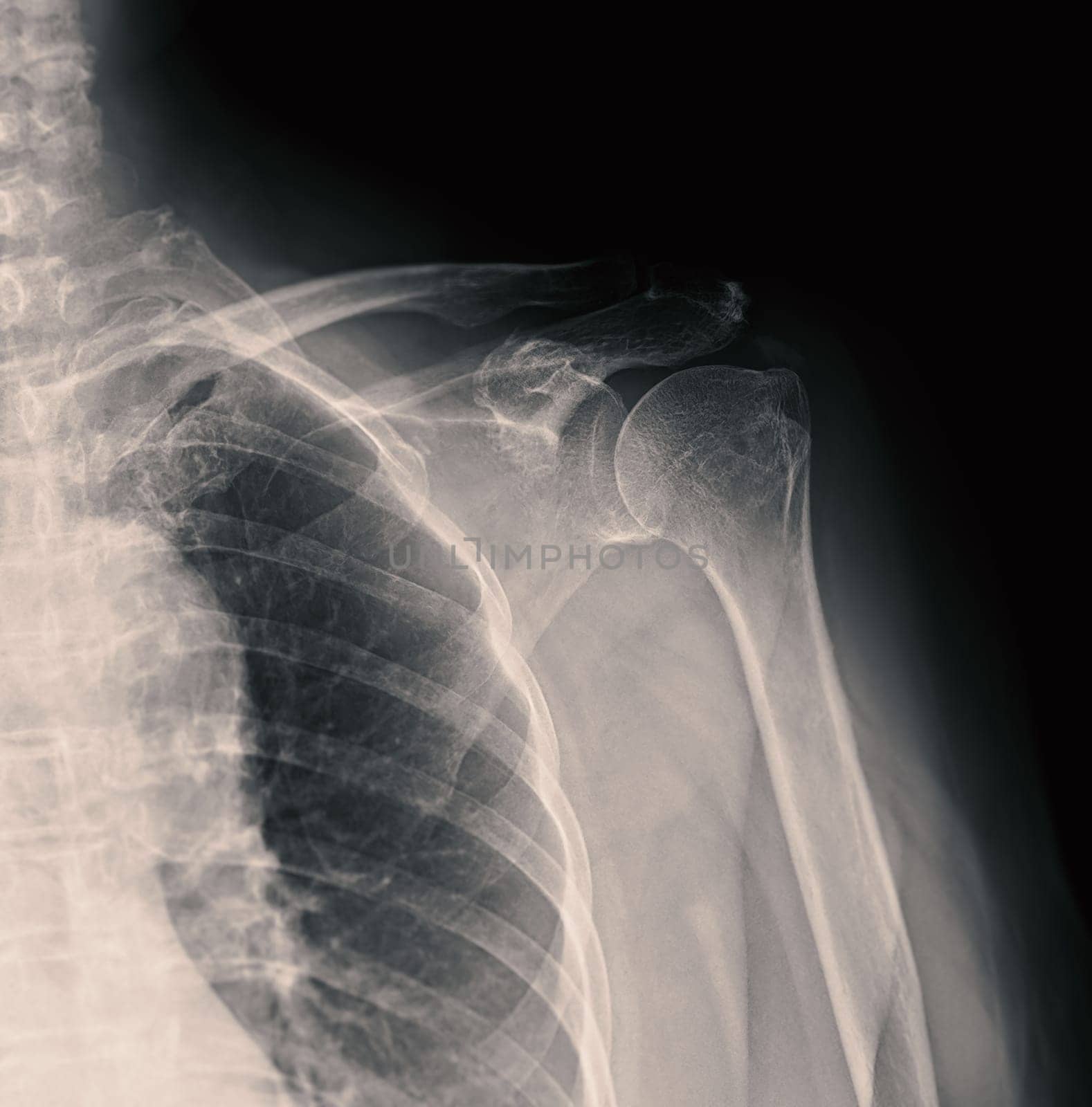
X-ray Shoulder joint shoulder front view for diagnosis fracture of shoulder joint.
Stock PhotoUsername
samunellaResolution
4626x4258pxX-ray Shoulder joint shoulder front view for diagnosis fracture of shoulder joint.

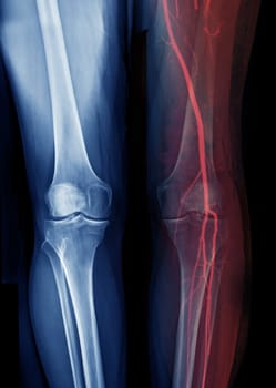
X-ray image of knee joint Fusion with CTA Femoral run off .
Stock PhotoUsername
samunellaResolution
4268x5992pxX-ray image of knee joint Fusion with CTA Femoral run off .
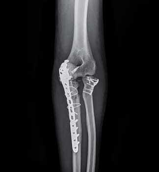
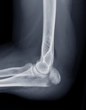
X-ray of Elbow join showing fracture of ulna bone.
Stock PhotoUsername
samunellaResolution
4920x3256pxX-ray of Elbow join showing fracture of ulna bone.

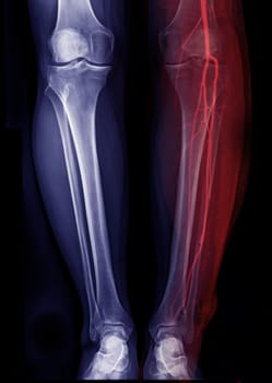
X-ray image of jeg Fusion with CTA Femoral run off .
Stock PhotoUsername
samunellaResolution
4268x5992pxX-ray image of jeg Fusion with CTA Femoral run off .
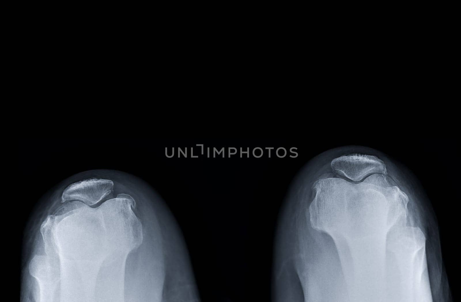
X-ray image of both Normal patella .
Stock PhotoUsername
samunellaResolution
6616x4332pxX-ray image of both Normal patella .

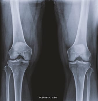
Film x-ray both knee joint AP view name is Rosenberg view for diagnosis knee pain from osteoarthritis knee and fracture .
Stock PhotoUsername
samunellaResolution
6590x6816pxFilm x-ray both knee joint AP view name is Rosenberg view for diagnosis knee pain from osteoarthritis knee and fracture .
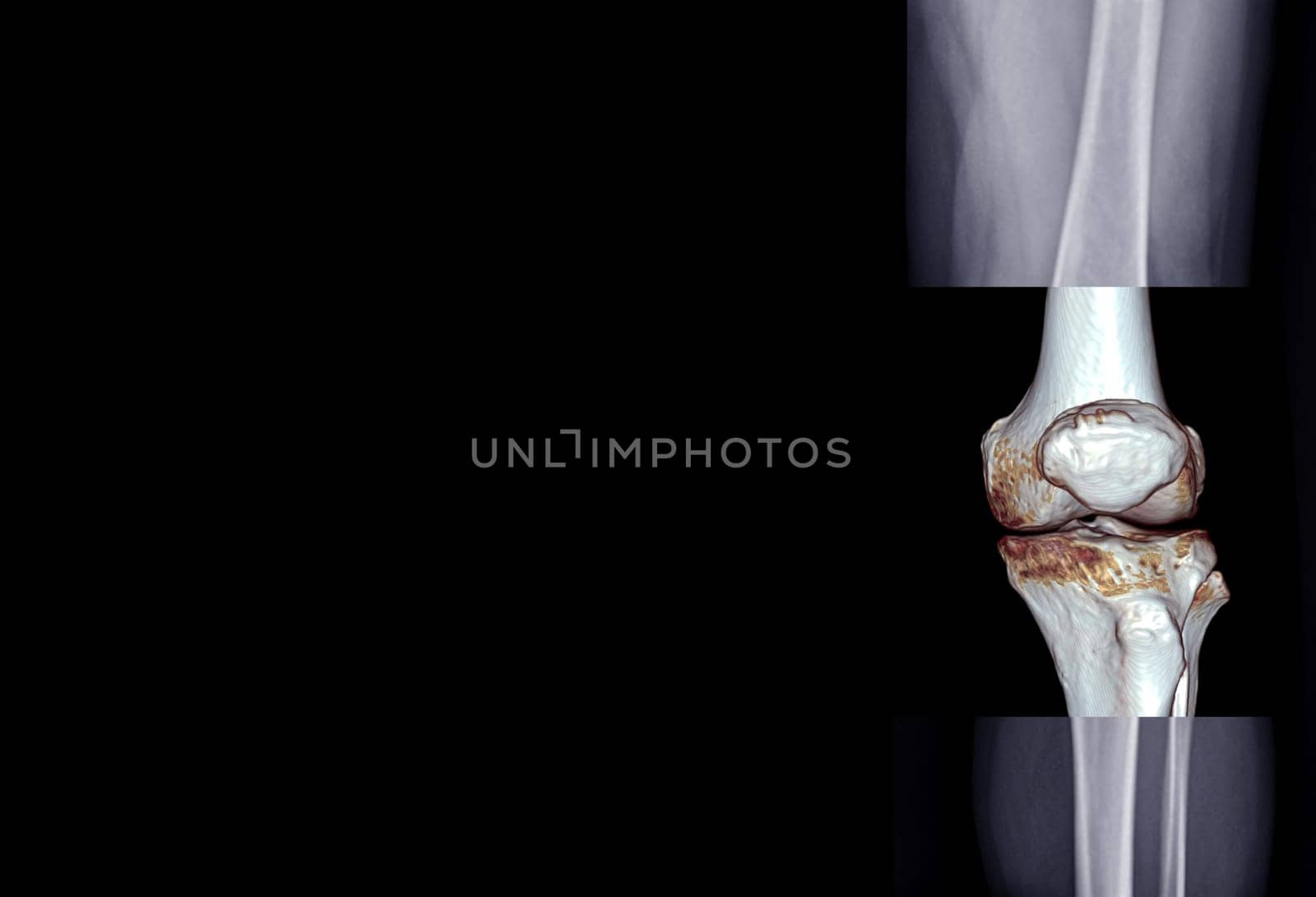
Film x-ray of knee joint AP view fusion with 3D rendering knee joint for medical background.
Stock PhotoUsername
samunellaResolution
9396x6400pxFilm x-ray of knee joint AP view fusion with 3D rendering knee joint for medical background.
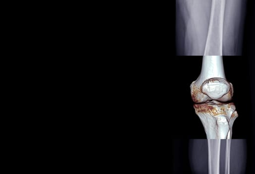

Film x-ray of knee joint AP view fusion with 3D rendering knee joint for medical background.
Stock PhotoUsername
samunellaResolution
9396x6400pxFilm x-ray of knee joint AP view fusion with 3D rendering knee joint for medical background.

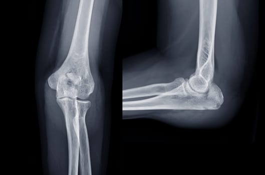
Film x-ray both knee joint AP view showing normal knee joint.
Stock PhotoUsername
samunellaResolution
6396x6400pxFilm x-ray both knee joint AP view showing normal knee joint.

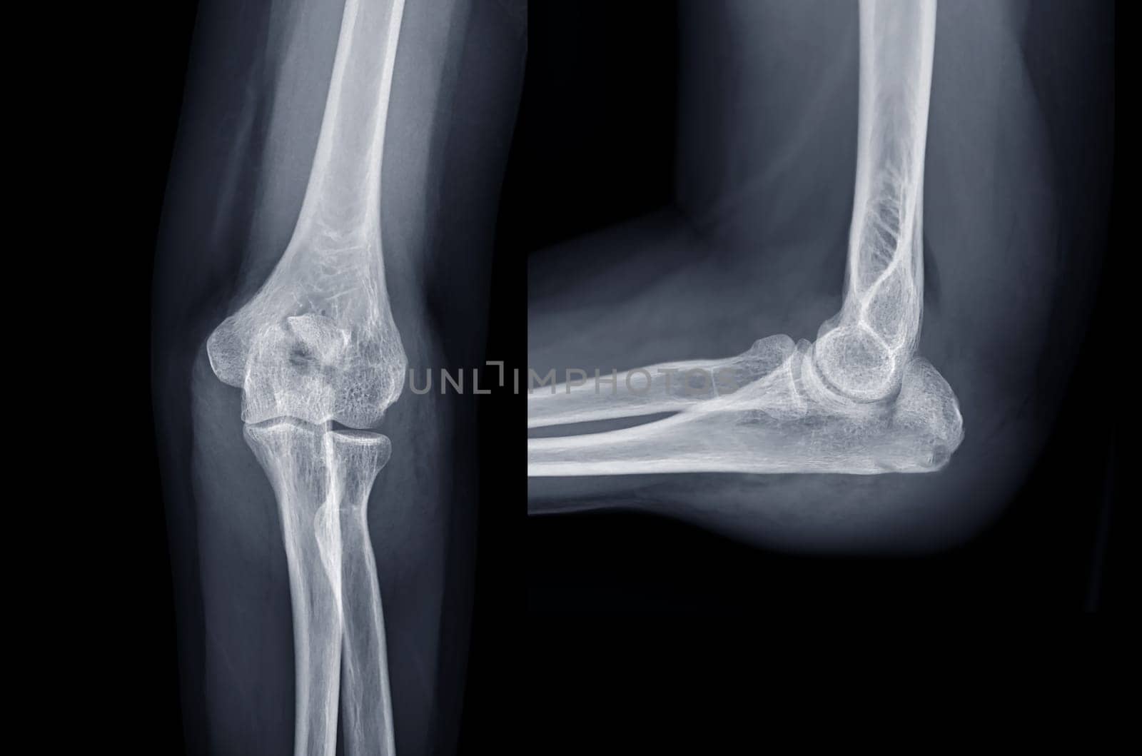
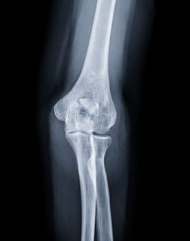
X-ray of Elbow join showing fracture of ulna bone.
Stock PhotoUsername
samunellaResolution
2618x3316pxX-ray of Elbow join showing fracture of ulna bone.

X-ray of Elbow join showing fracture of ulna bone.
Stock PhotoUsername
samunellaResolution
2618x3316pxX-ray of Elbow join showing fracture of ulna bone.
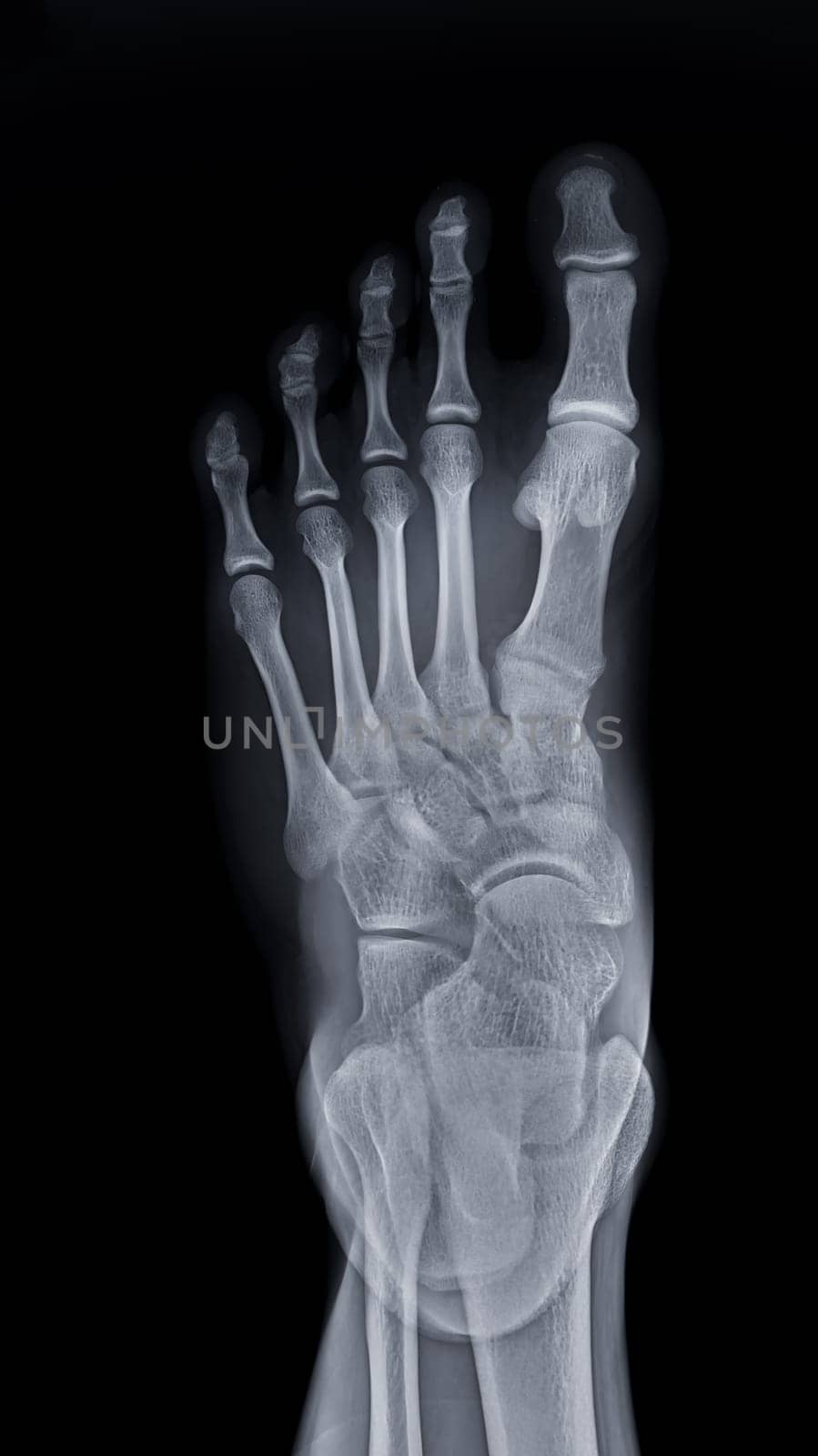
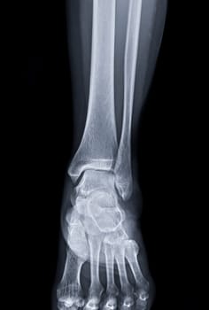
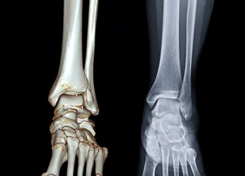
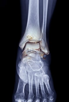
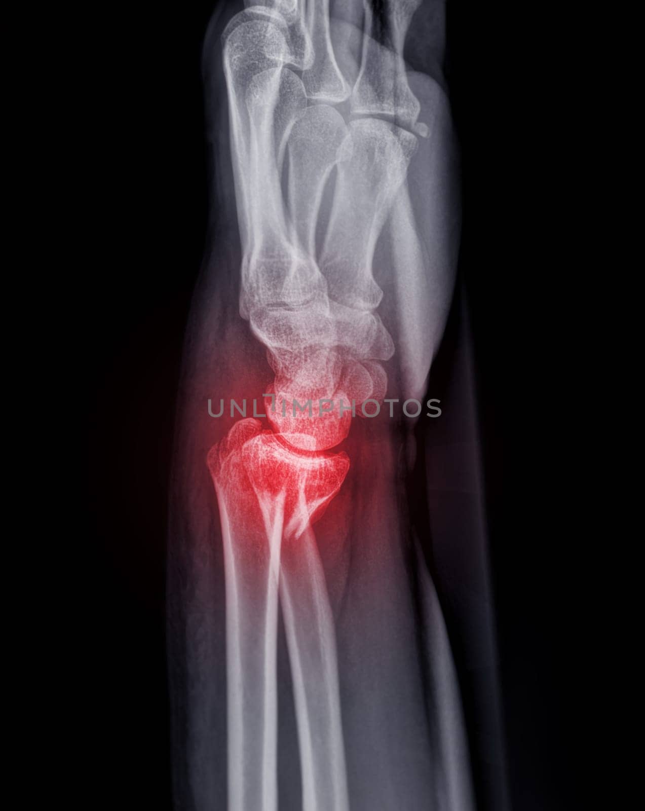
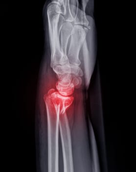
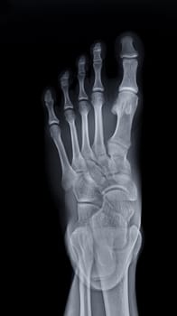
Foot x-ray image AP view isolated on black background.
Stock PhotoUsername
samunellaResolution
2744x4882pxFoot x-ray image AP view isolated on black background.

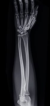
X-ray image of forearm bone.
Stock PhotoUsername
samunellaResolution
3094x6464pxX-ray image of forearm bone.
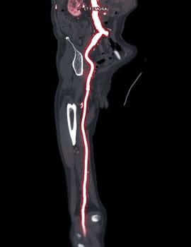
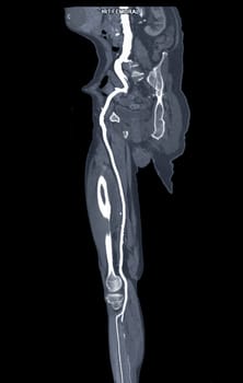
CTA femoral artery run off MPR curve showing Right femoral artery for diagnostic Acute or Chronic Peripheral Arterial Disease.
Stock PhotoUsername
samunellaResolution
2794x4410pxCTA femoral artery run off MPR curve showing Right femoral artery for diagnostic Acute or Chronic Peripheral Arterial Disease.
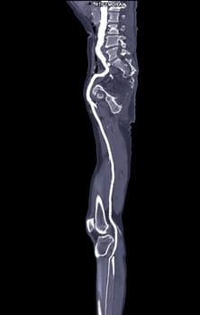

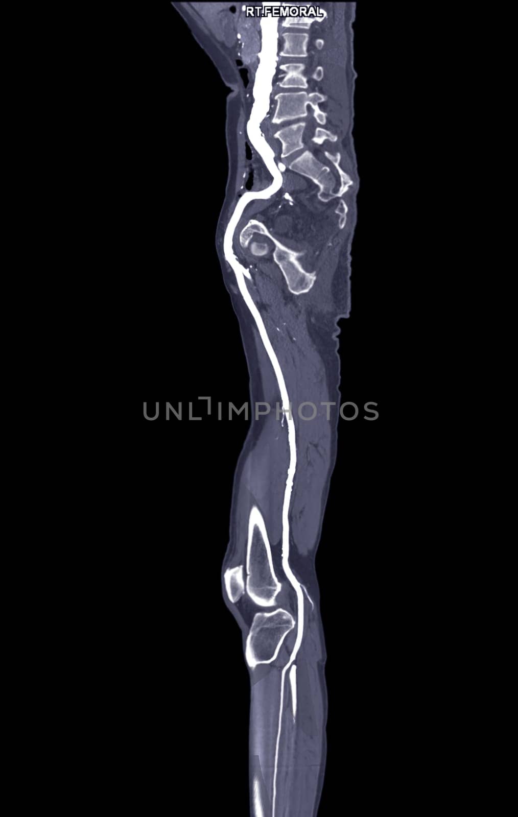
CTA femoral artery run off MPR curve showing Left femoral artery for diagnostic Acute or Chronic Peripheral Arterial Disease.
Stock PhotoUsername
samunellaResolution
2659x3438pxCTA femoral artery run off MPR curve showing Left femoral artery for diagnostic Acute or Chronic Peripheral Arterial Disease.
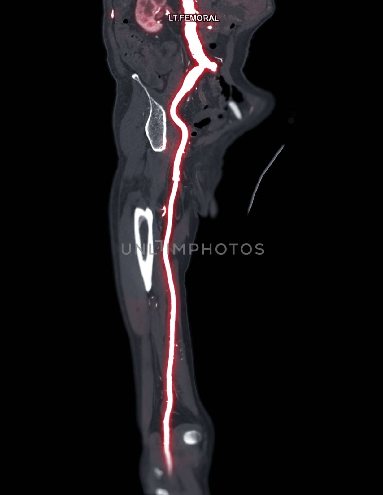