- Filter By:
-
-
Stock photos and images of username:samunella

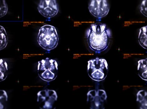
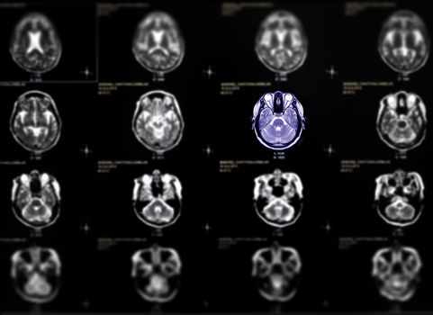
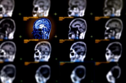
Selective focus of MRI brain axial view for detect a variety of conditions of the brain stroke disease.
Stock PhotoUsername
samunellaResolution
6090x4514pxSelective focus of MRI brain axial view for detect a variety of conditions of the brain stroke disease.

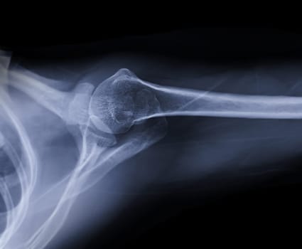
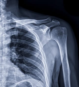
X-ray Shoulder joint shoulder transaxillary view for diagnosis fracture of shoulder joint.
Stock PhotoUsername
samunellaResolution
3678x3033pxX-ray Shoulder joint shoulder transaxillary view for diagnosis fracture of shoulder joint.
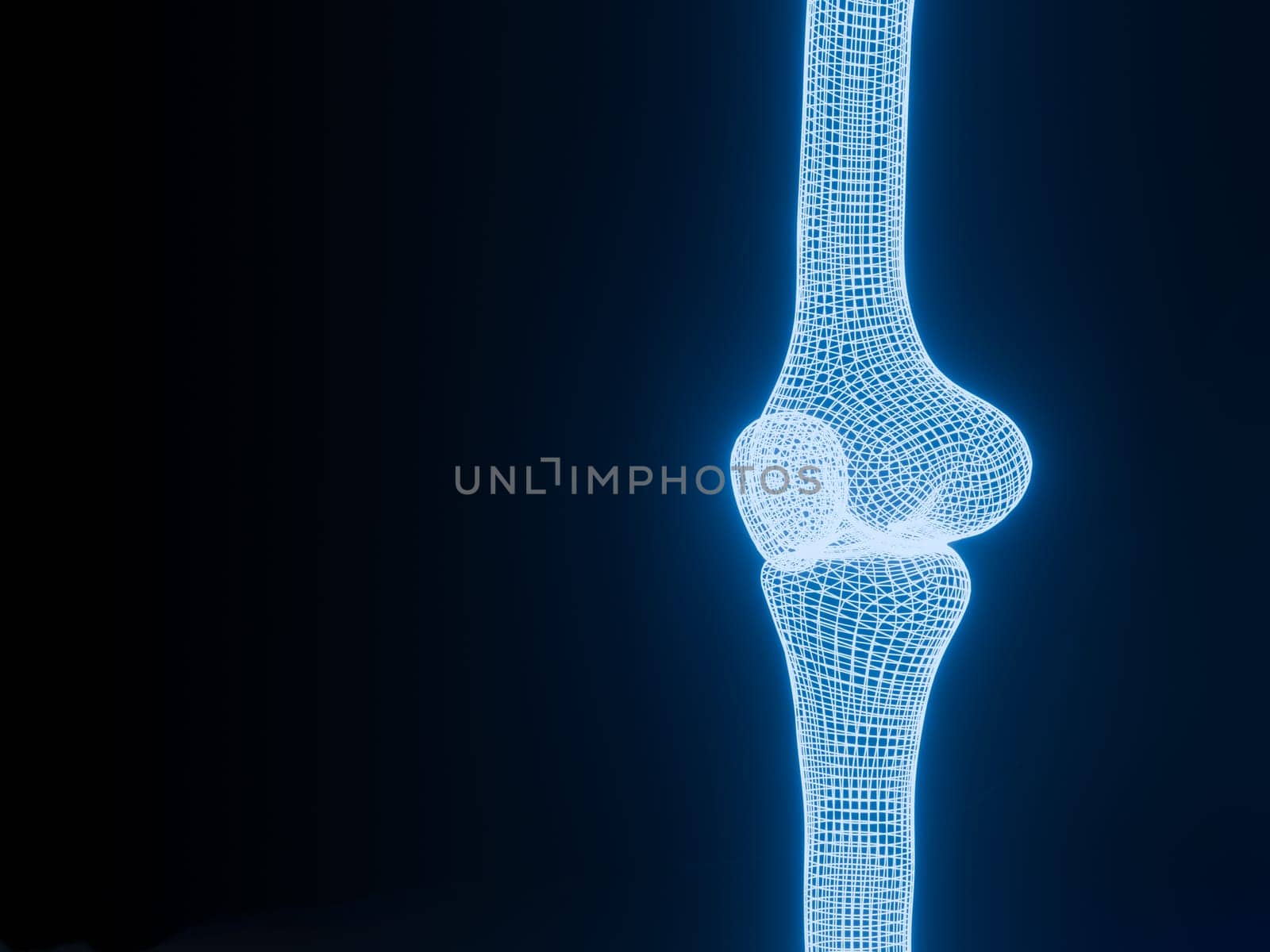
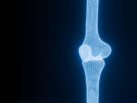
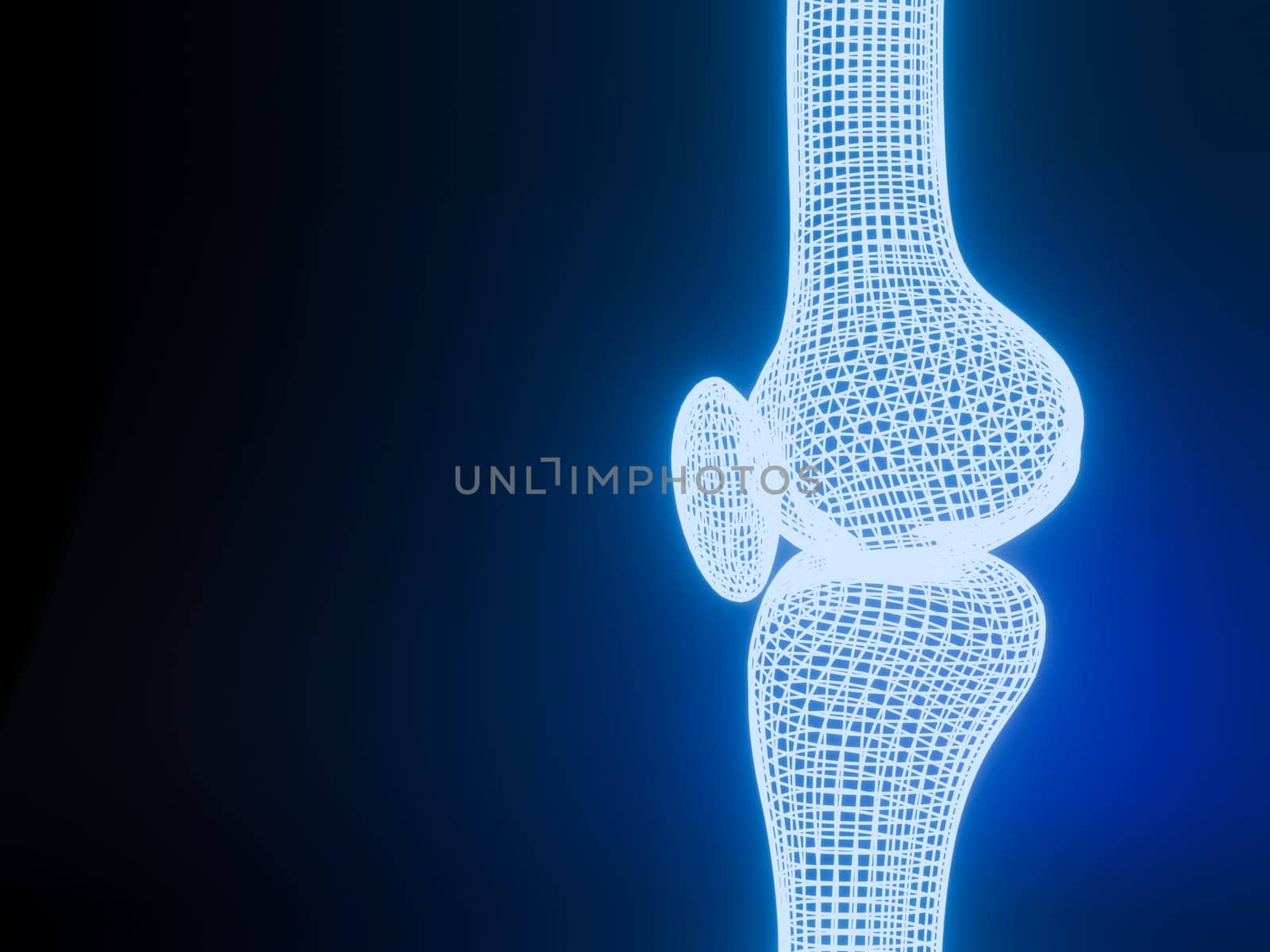
Knee joint 3D rendering Blue glowing wireframe on black background with copy space for text. Bone human skeleton anatomy of the body. Medical health care science concept. Realistic 3D Rendering.
Stock PhotoUsername
samunellaResolution
4000x3000pxKnee joint 3D rendering Blue glowing wireframe on black background with copy space for text. Bone human skeleton anatomy of the body. Medical health care science concept. Realistic 3D Rendering.

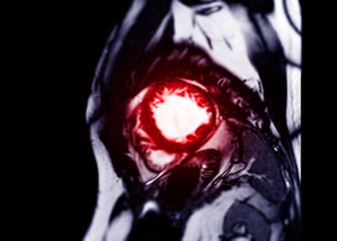
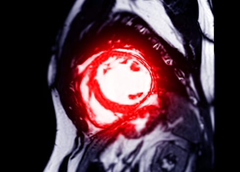
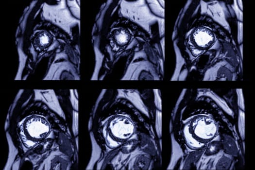
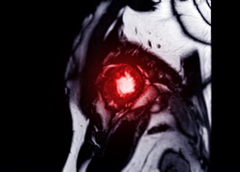
MRI heart or Cardiac MRI in short axis view showing cross-sections of the left and right ventricle for diagnosis heart disease.
Stock PhotoUsername
samunellaResolution
3430x2458pxMRI heart or Cardiac MRI in short axis view showing cross-sections of the left and right ventricle for diagnosis heart disease.
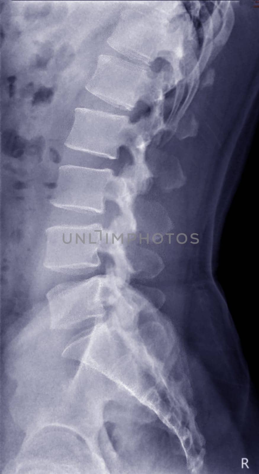
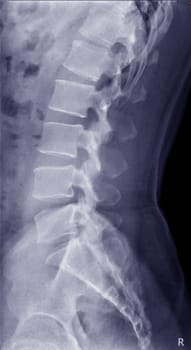
X-ray image of lambosacral spine or L-S spine
Stock PhotoUsername
samunellaResolution
2467x4512pxX-ray image of lambosacral spine or L-S spine

milkyway rise above sea with waves trails for background.
Stock PhotoUsername
samunellaResolution
5739x3826pxmilkyway rise above sea with waves trails for background.


Selective focus of MRI brain axial view for detect a variety of conditions of the brain stroke disease.
Stock PhotoUsername
samunellaResolution
7408x5392pxSelective focus of MRI brain axial view for detect a variety of conditions of the brain stroke disease.

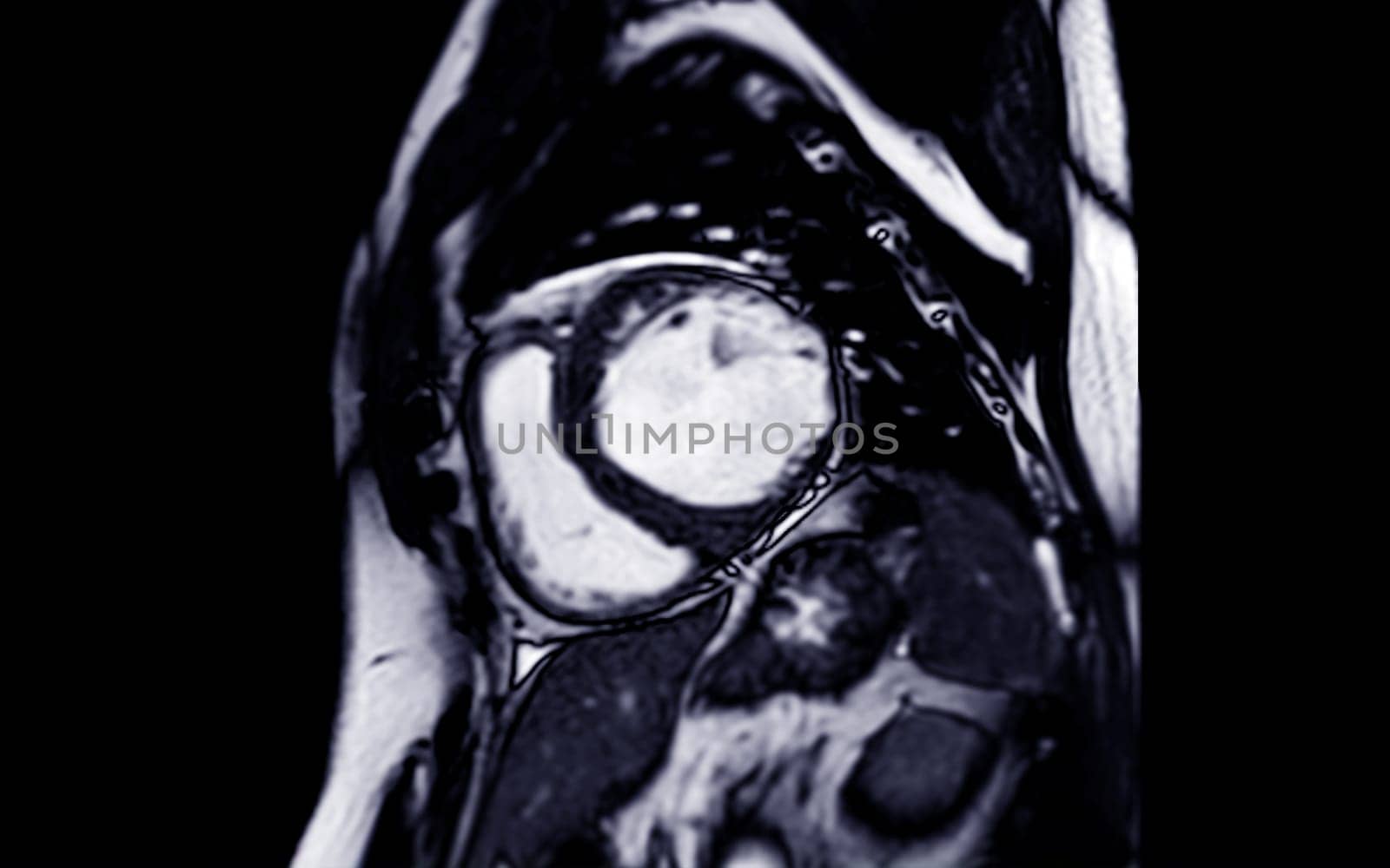
MRI heart or Cardiac MRI in short axis view showing cross-sections of the left and right ventricle for diagnosis heart disease.
Stock PhotoUsername
samunellaResolution
3430x2458pxMRI heart or Cardiac MRI in short axis view showing cross-sections of the left and right ventricle for diagnosis heart disease.

X-ray of Shoulder joint for diagnosis shoulder joint from dislocation or fracture.
Stock PhotoUsername
samunellaResolution
4160x4556pxX-ray of Shoulder joint for diagnosis shoulder joint from dislocation or fracture.
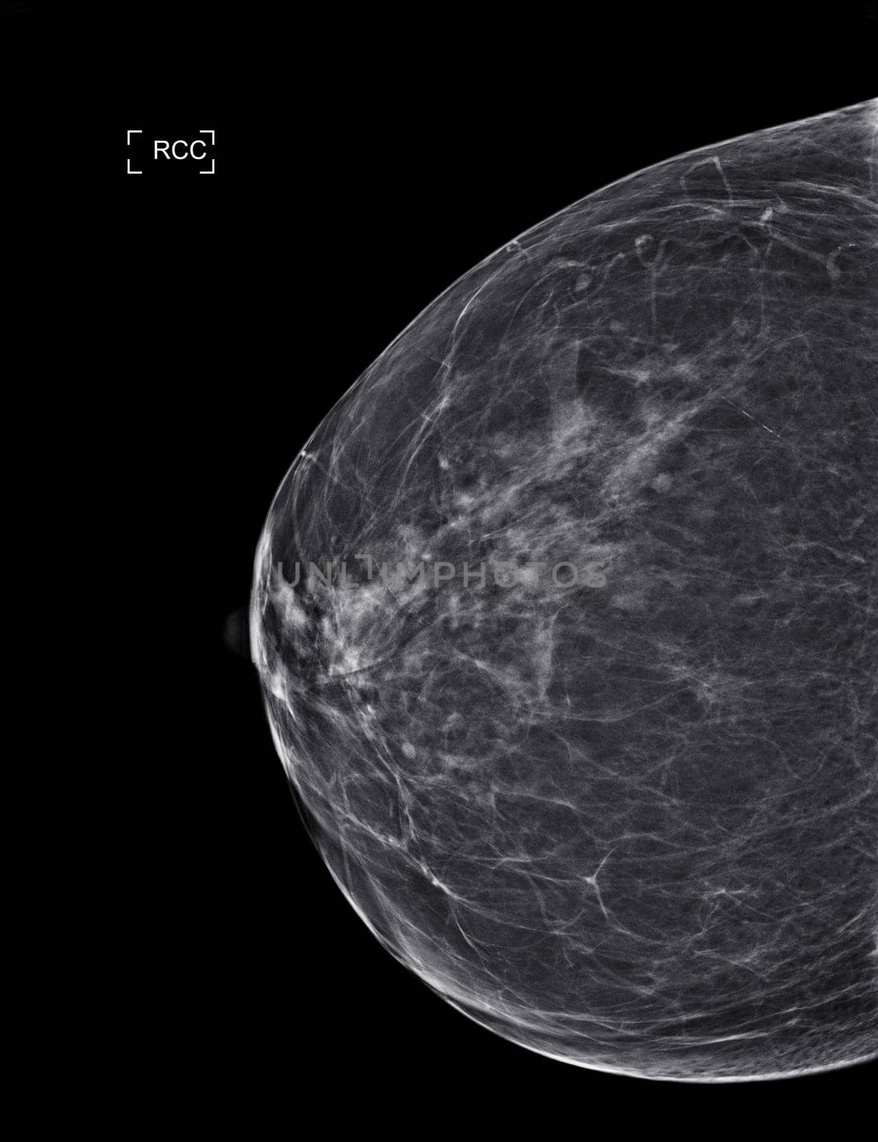
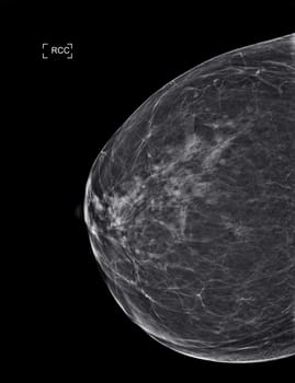
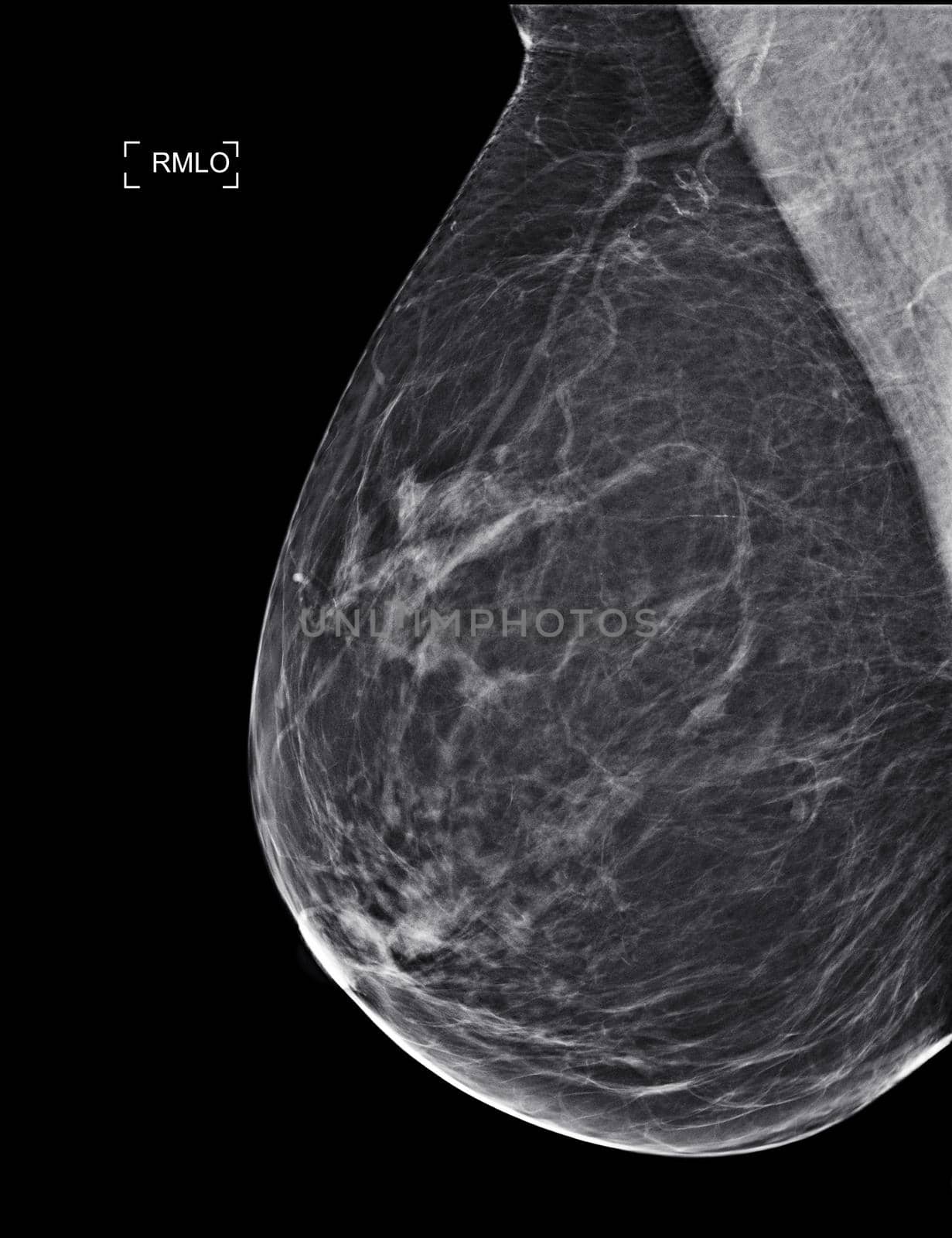
X-ray Digital Mammogram or mammography
Stock PhotoUsername
samunellaResolution
2560x3328pxX-ray Digital Mammogram or mammography

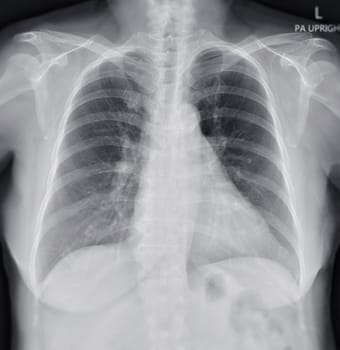
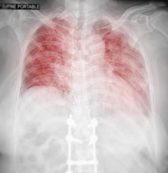
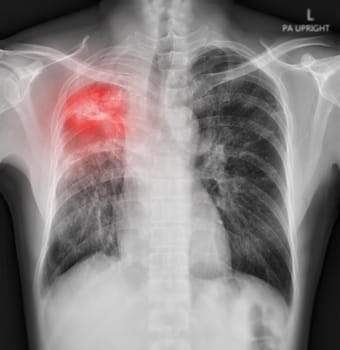
Chest x-ray image for screening diagnosis TB,tuberculosis and covid-19.
Stock PhotoUsername
samunellaResolution
2701x2784pxChest x-ray image for screening diagnosis TB,tuberculosis and covid-19.

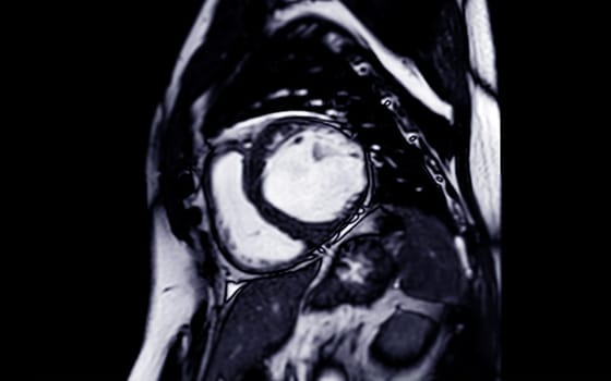
MRI heart or Cardiac MRI in short axis view showing cross-sections of the left and right ventricle for diagnosis heart disease.
Stock PhotoUsername
samunellaResolution
6000x4000pxMRI heart or Cardiac MRI in short axis view showing cross-sections of the left and right ventricle for diagnosis heart disease.

Chest x-ray image for screening diagnosis TB,tuberculosis and covid-19.
Stock PhotoUsername
samunellaResolution
2701x2784pxChest x-ray image for screening diagnosis TB,tuberculosis and covid-19.
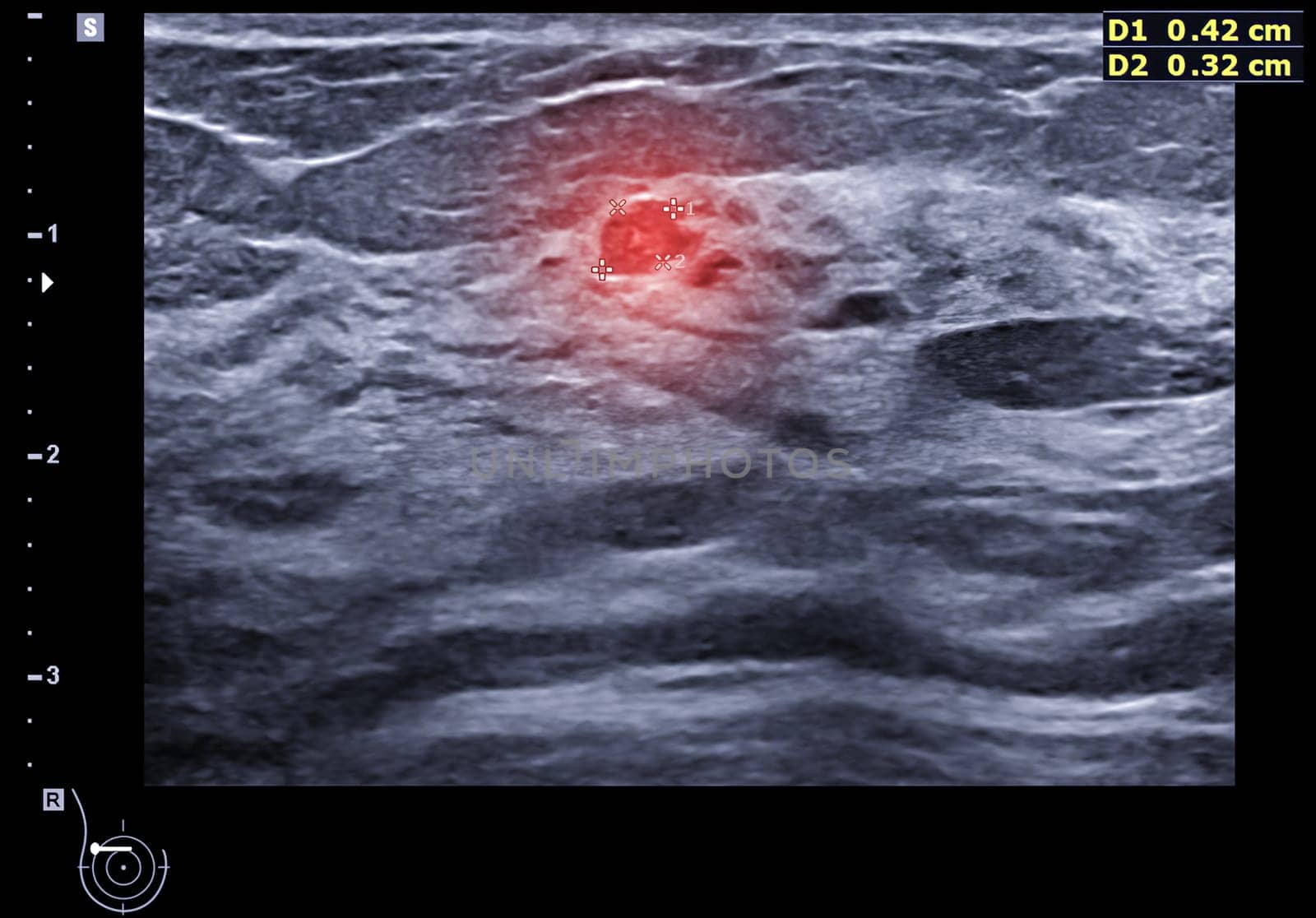
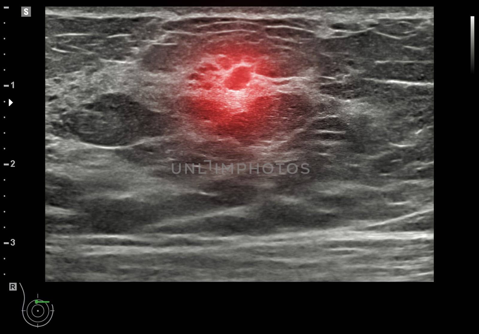
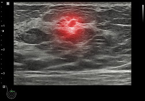
ultrasound breast of Patient after mammogram for diagnonsis Breast cancer in women isolated on black background.
Stock PhotoUsername
samunellaResolution
3096x2160pxultrasound breast of Patient after mammogram for diagnonsis Breast cancer in women isolated on black background.

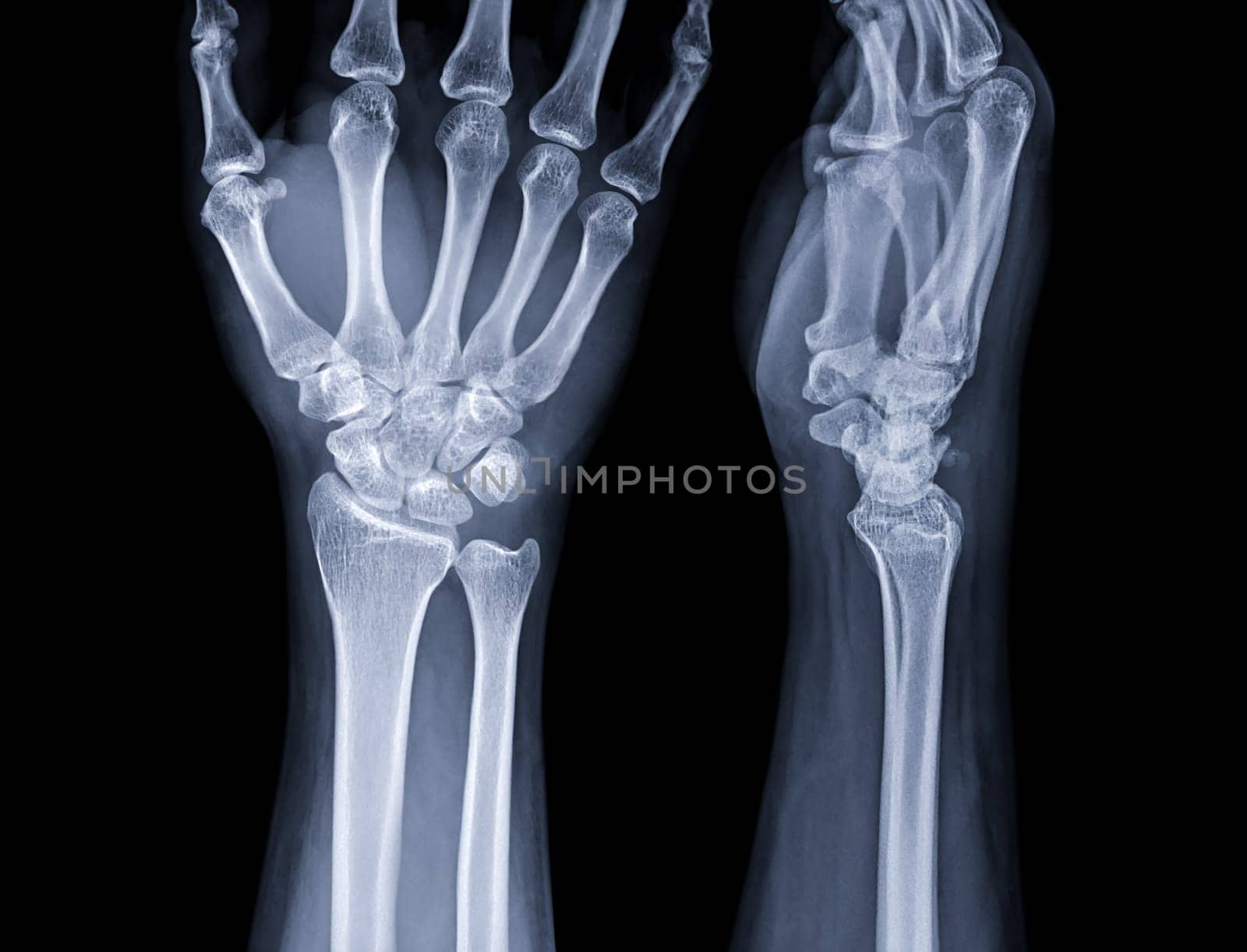
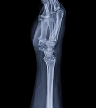
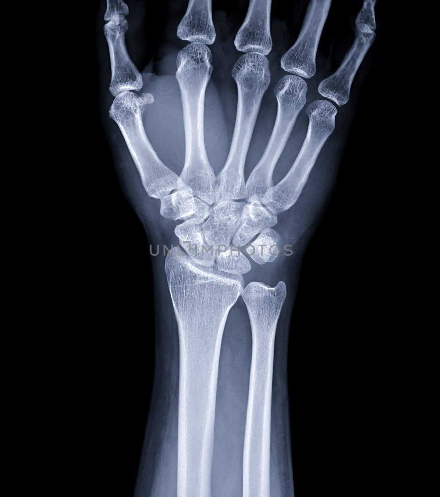
X-ray image of wrist joint for diagnosis rheumatoid arthritis .
Stock PhotoUsername
samunellaResolution
5276x4026pxX-ray image of wrist joint for diagnosis rheumatoid arthritis .

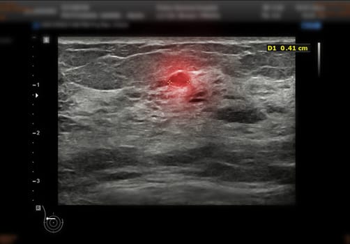
ultrasound breast of Patient after mammogram for diagnonsis Breast cancer in women isolated on black background.
Stock PhotoUsername
samunellaResolution
3096x2160pxultrasound breast of Patient after mammogram for diagnonsis Breast cancer in women isolated on black background.

MRI heart or Cardiac MRI in short axis view showing cross-sections of the left and right ventricle for diagnosis heart disease.
Stock PhotoUsername
samunellaResolution
3430x2458pxMRI heart or Cardiac MRI in short axis view showing cross-sections of the left and right ventricle for diagnosis heart disease.
MRI heart or Cardiac MRI in short axis view showing cross-sections of the left and right ventricle for diagnosis heart disease.
Stock PhotoUsername
samunellaResolution
3840x2400pxMRI heart or Cardiac MRI in short axis view showing cross-sections of the left and right ventricle for diagnosis heart disease.
Knee joint 3D rendering Blue glowing wireframe on black background with copy space for text. Bone human skeleton anatomy of the body. Medical health care science concept. Realistic 3D Rendering.
Stock PhotoUsername
samunellaResolution
4000x3000pxKnee joint 3D rendering Blue glowing wireframe on black background with copy space for text. Bone human skeleton anatomy of the body. Medical health care science concept. Realistic 3D Rendering.

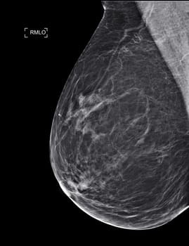
X-ray Digital Mammogram or mammography
Stock PhotoUsername
samunellaResolution
2560x3328pxX-ray Digital Mammogram or mammography

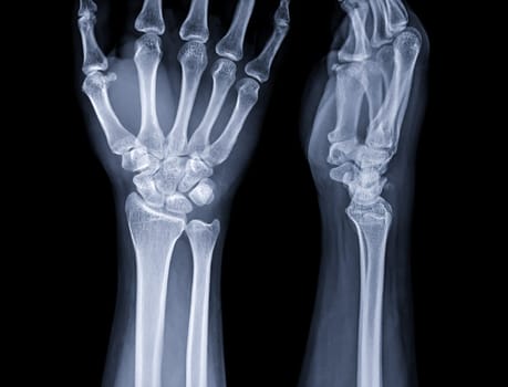
X-ray image of wrist joint for diagnosis rheumatoid arthritis .
Stock PhotoUsername
samunellaResolution
3564x4026pxX-ray image of wrist joint for diagnosis rheumatoid arthritis .
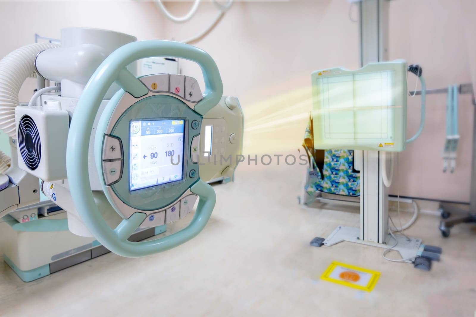
X-ray generator tube with monitor or X-ray general device with Bucky stand in radiology .modern medical equipment in the hospital.
Stock PhotoUsername
samunellaResolution
6000x4000pxX-ray generator tube with monitor or X-ray general device with Bucky stand in radiology .modern medical equipment in the hospital.

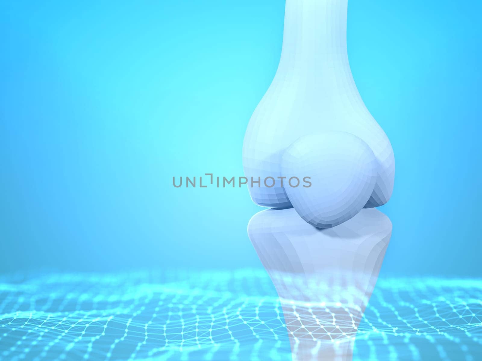
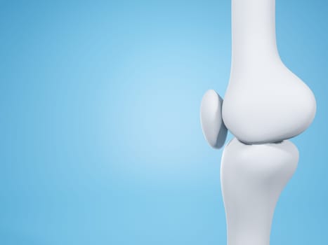
Knee joint on blue background with copy space for text. Bone human skeleton anatomy of the body. Medical health care science concept. Realistic 3D Rendering.
Stock PhotoUsername
samunellaResolution
4000x3000pxKnee joint on blue background with copy space for text. Bone human skeleton anatomy of the body. Medical health care science concept. Realistic 3D Rendering.

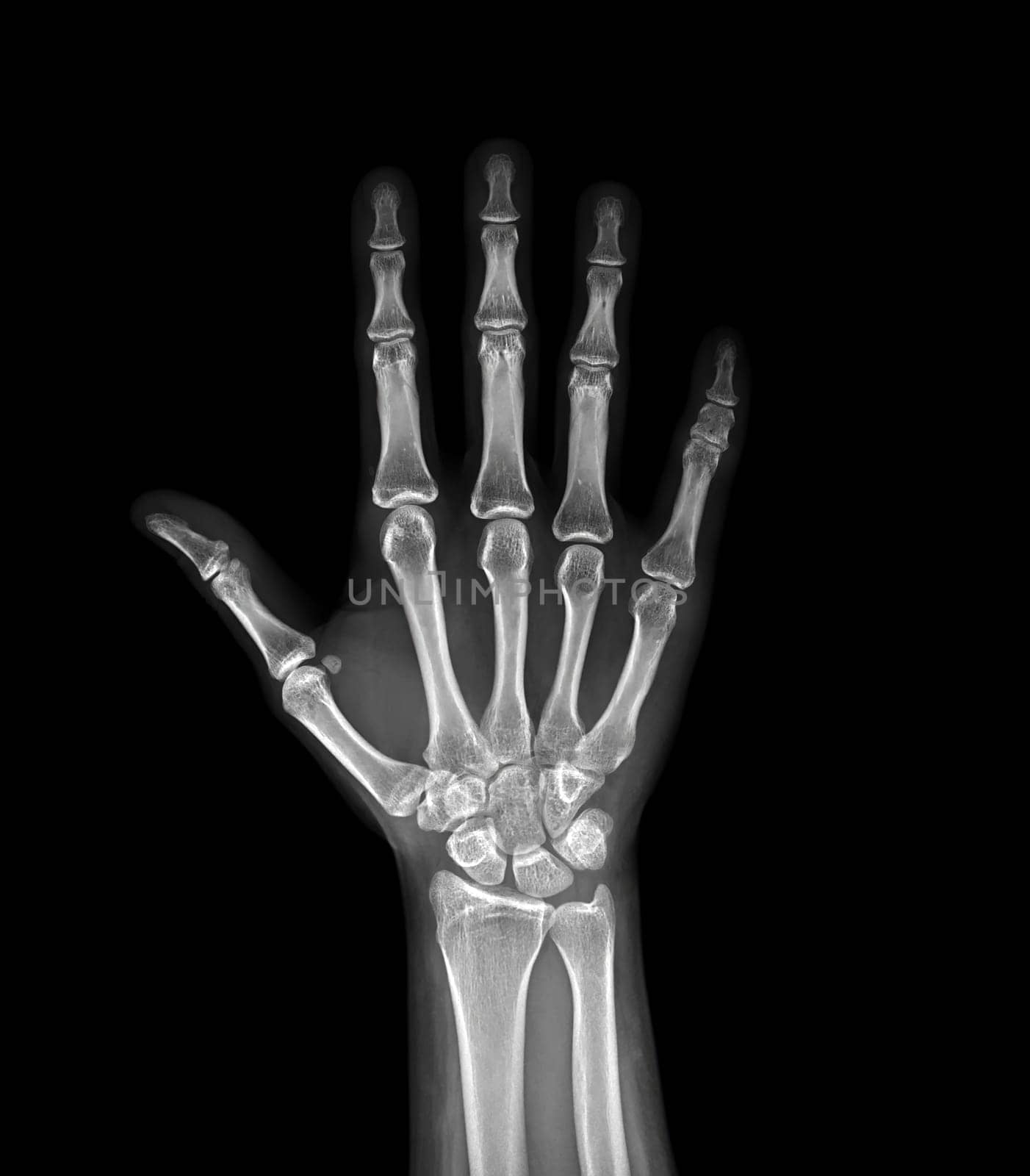
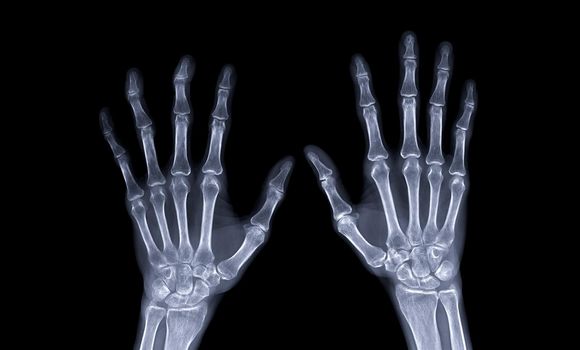
xray image of both hand AP view isolated on black background for diagnostic rheumatoid.
Stock PhotoUsername
samunellaResolution
5150x5876pxxray image of both hand AP view isolated on black background for diagnostic rheumatoid.
ultrasound breast of Patient after mammogram for diagnonsis Breast cancer in women isolated on black background.
Stock PhotoUsername
samunellaResolution
3096x2160pxultrasound breast of Patient after mammogram for diagnonsis Breast cancer in women isolated on black background.
X-ray image of wrist joint for diagnosis rheumatoid arthritis .
Stock PhotoUsername
samunellaResolution
3564x4026pxX-ray image of wrist joint for diagnosis rheumatoid arthritis .


Chest x-ray image for screening diagnosis TB,tuberculosis and covid-19.
Stock PhotoUsername
samunellaResolution
2701x2784pxChest x-ray image for screening diagnosis TB,tuberculosis and covid-19.

Selective focus of MRI brain axial view for detect a variety of conditions of the brain stroke disease.
Stock PhotoUsername
samunellaResolution
7168x4728pxSelective focus of MRI brain axial view for detect a variety of conditions of the brain stroke disease.

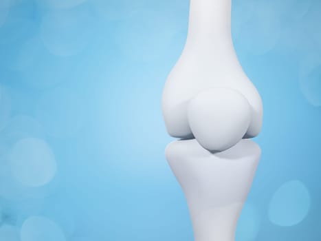
Knee joint on blue background with copy space for text. Bone human skeleton anatomy of the body. Medical health care science concept. Realistic 3D Rendering.
Stock PhotoUsername
samunellaResolution
4000x3000pxKnee joint on blue background with copy space for text. Bone human skeleton anatomy of the body. Medical health care science concept. Realistic 3D Rendering.

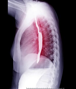
Esophagram or Barium swallow
Stock PhotoUsername
samunellaResolution
2417x2816pxEsophagram or Barium swallow

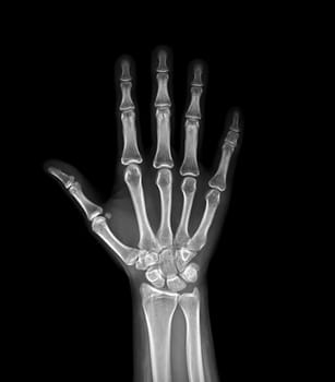
xray image of both hand AP view isolated on black background for diagnostic rheumatoid.
Stock PhotoUsername
samunellaResolution
7416x4472pxxray image of both hand AP view isolated on black background for diagnostic rheumatoid.
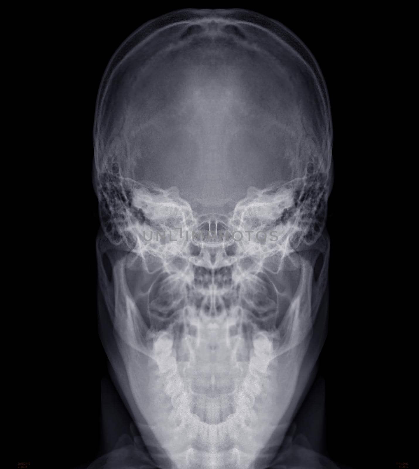
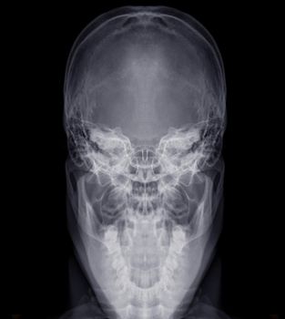
Skull x-ray image of Human name is skull towne's view .isolated on Black Background.
Stock PhotoUsername
samunellaResolution
2640x2952pxSkull x-ray image of Human name is skull towne's view .isolated on Black Background.
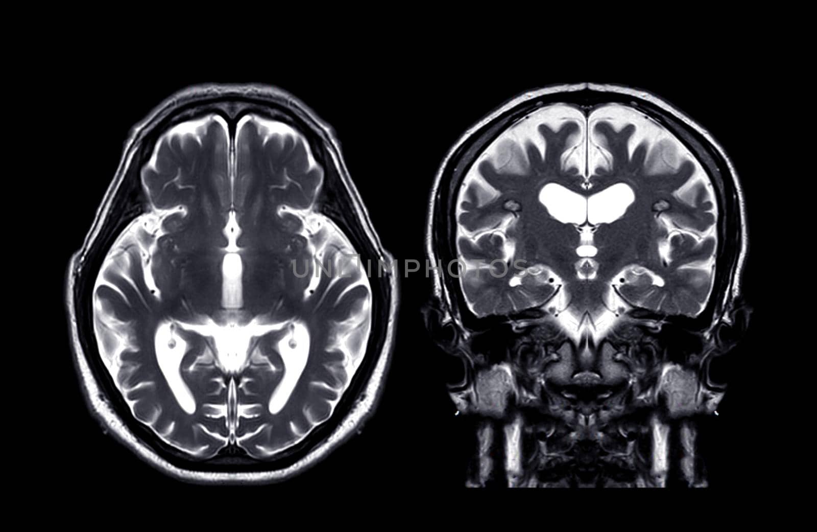
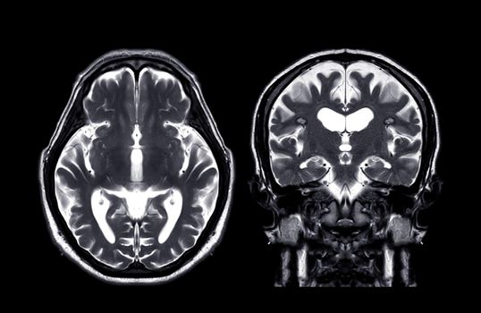
MRI brain Axial T2 and coronal t2 f technique
Stock PhotoUsername
samunellaResolution
4583x2983pxMRI brain Axial T2 and coronal t2 f technique

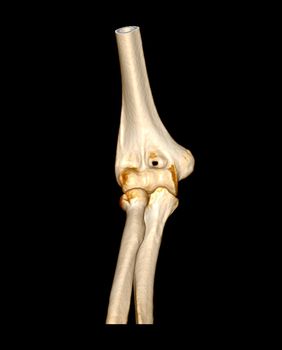
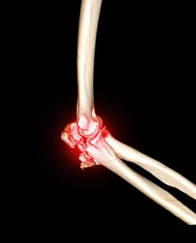
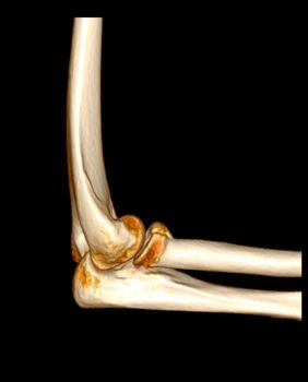
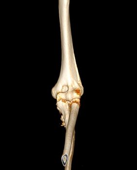
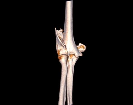
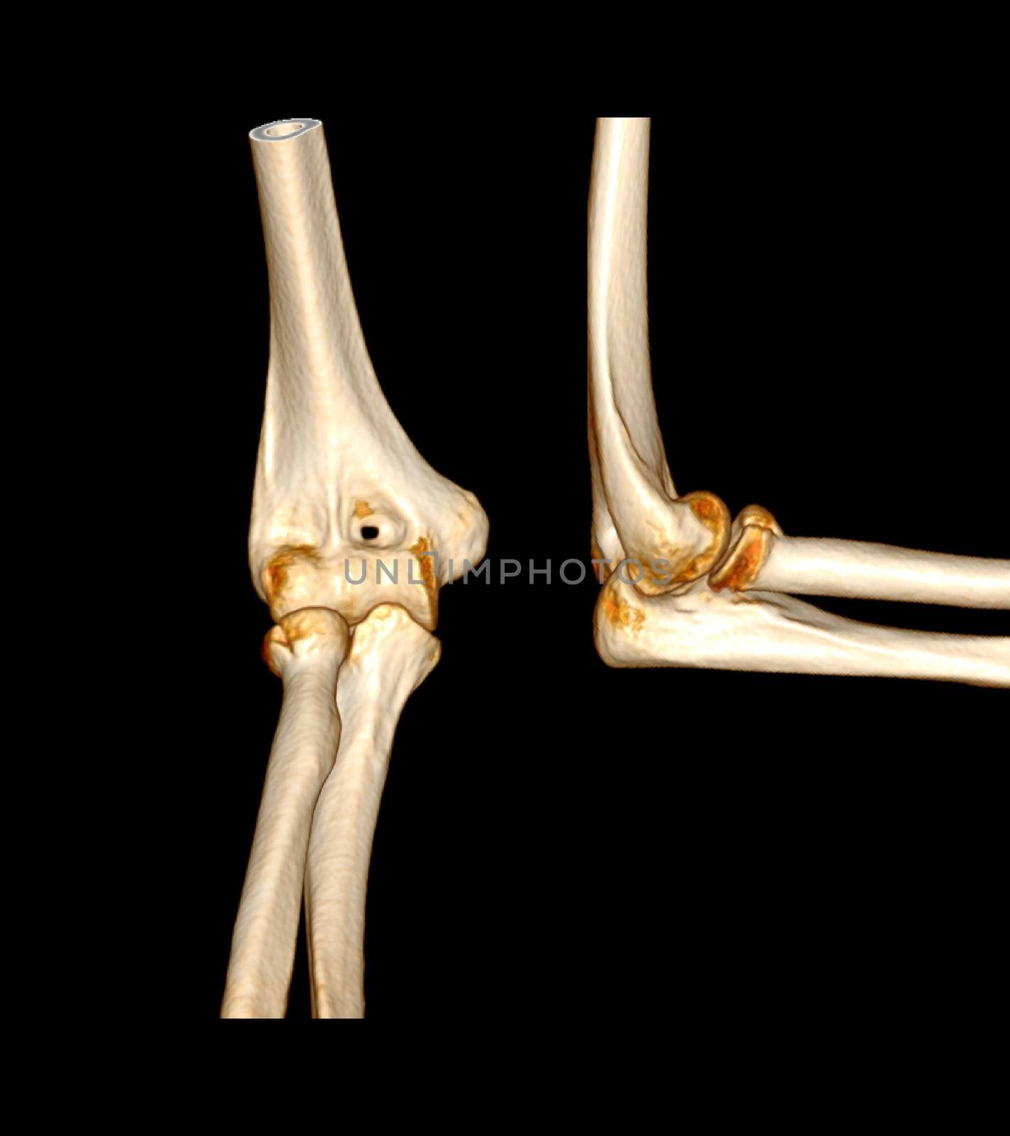
CT scan of elbow joint
Stock PhotoUsername
samunellaResolution
2542x3154pxCT scan of elbow joint
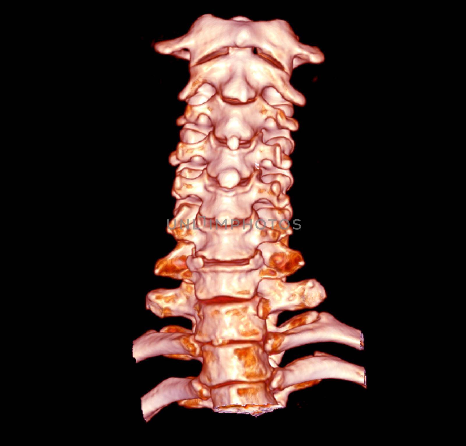
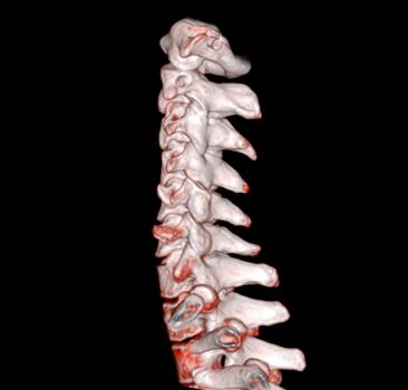
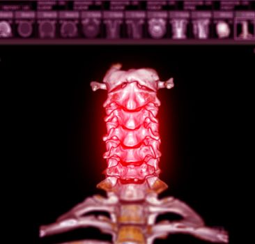
CT SCAN of Cervical Spine 3D rendering .
Stock PhotoUsername
samunellaResolution
3344x3200pxCT SCAN of Cervical Spine 3D rendering .

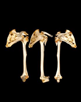
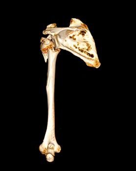
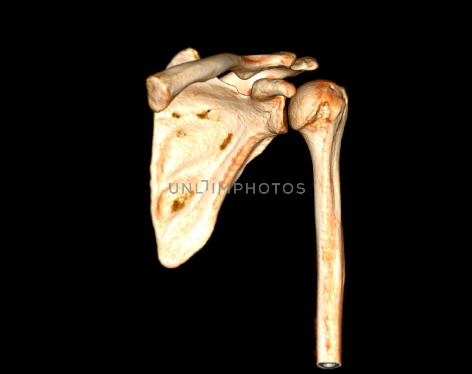
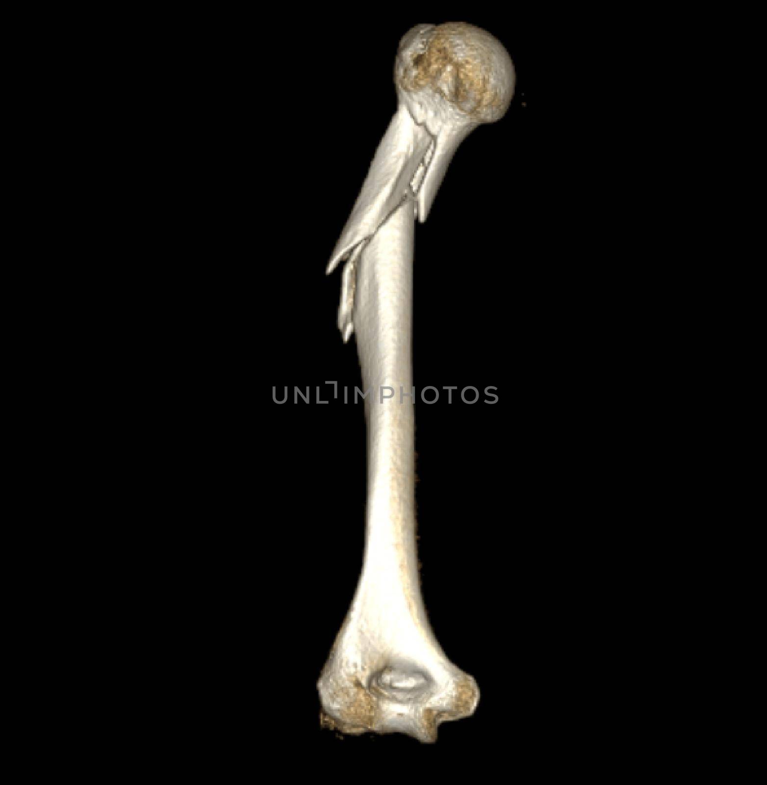
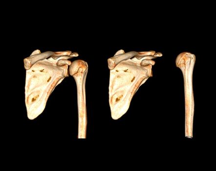
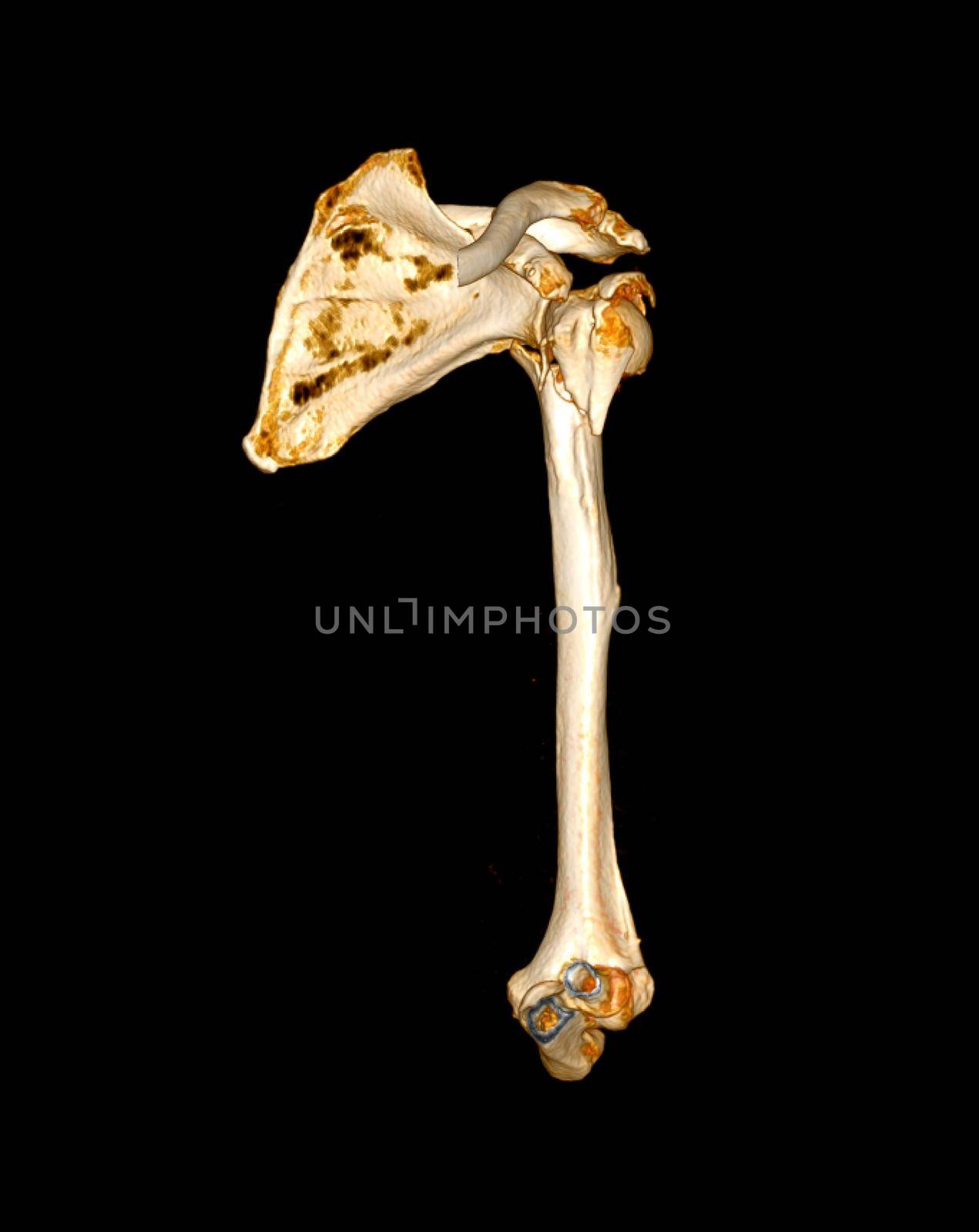
CT scan of shoulder joint and humerus bone or arm.
Stock PhotoUsername
samunellaResolution
2894x3642pxCT scan of shoulder joint and humerus bone or arm.

CT scan of elbow joint
Stock PhotoUsername
samunellaResolution
2542x3154pxCT scan of elbow joint
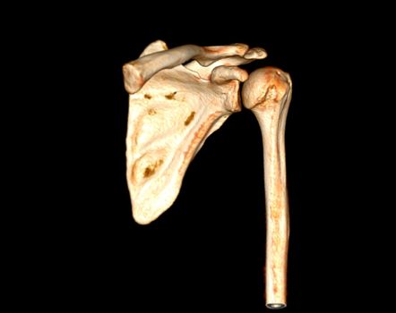
CT scan of shoulder joint.
Stock PhotoUsername
samunellaResolution
4040x3200pxCT scan of shoulder joint.

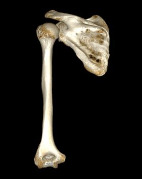
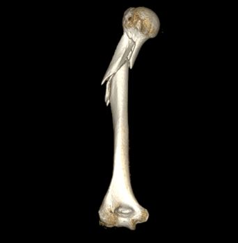
CT scan of shoulder joint and humerus bone or arm.
Stock PhotoUsername
samunellaResolution
2894x3642pxCT scan of shoulder joint and humerus bone or arm.

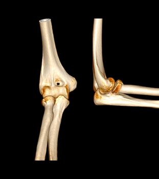
CT scan of elbow joint
Stock PhotoUsername
samunellaResolution
2542x3154pxCT scan of elbow joint
CT scan of shoulder joint and humerus bone or arm.
Stock PhotoUsername
samunellaResolution
3128x3200pxCT scan of shoulder joint and humerus bone or arm.

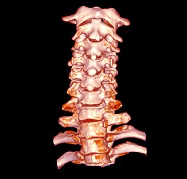
CT SCAN of Cervical Spine 3D rendering .
Stock PhotoUsername
samunellaResolution
3344x3200pxCT SCAN of Cervical Spine 3D rendering .

CT scan of elbow joint
Stock PhotoUsername
samunellaResolution
2542x3154pxCT scan of elbow joint

CT scan of shoulder joint.
Stock PhotoUsername
samunellaResolution
4040x3200pxCT scan of shoulder joint.

CT scan of elbow joint
Stock PhotoUsername
samunellaResolution
4040x3200pxCT scan of elbow joint

CT SCAN of Cervical Spine 3D rendering .
Stock PhotoUsername
samunellaResolution
3344x3200pxCT SCAN of Cervical Spine 3D rendering .
CT scan of elbow joint
Stock PhotoUsername
samunellaResolution
2806x3154pxCT scan of elbow joint
CT scan of shoulder joint and humerus bone or arm.
Stock PhotoUsername
samunellaResolution
2894x3642pxCT scan of shoulder joint and humerus bone or arm.


CT scan of shoulder joint and humerus bone or arm.
Stock PhotoUsername
samunellaResolution
2894x3642pxCT scan of shoulder joint and humerus bone or arm.