- Filter By:
-
-
Stock photos and images of username:creativepic

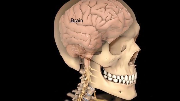
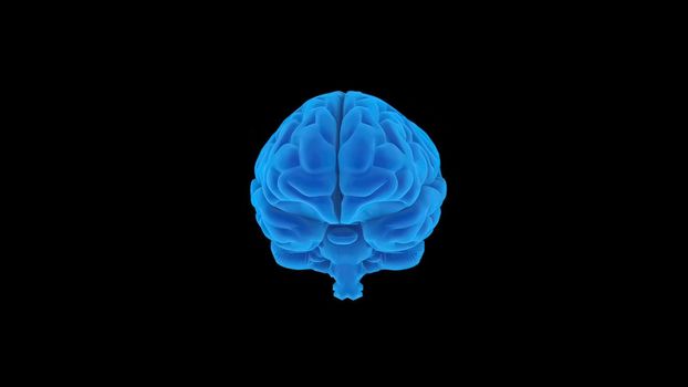
3D human brain model from external on black ackground
Stock PhotoUsername
creativepicResolution
3840x2160px3D human brain model from external on black ackground

Human Gall Bladder Anatomy With Digestive System For Medical Concept
Stock PhotoUsername
creativepicResolution
3840x2160pxHuman Gall Bladder Anatomy With Digestive System For Medical Concept
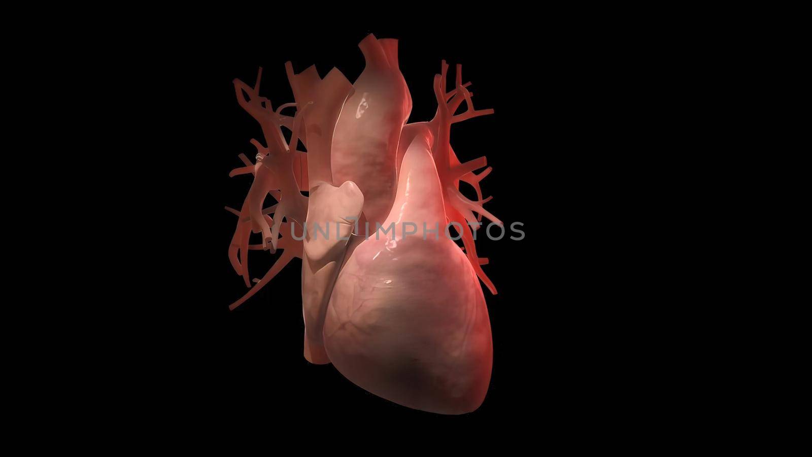
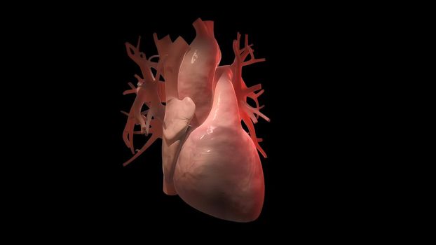
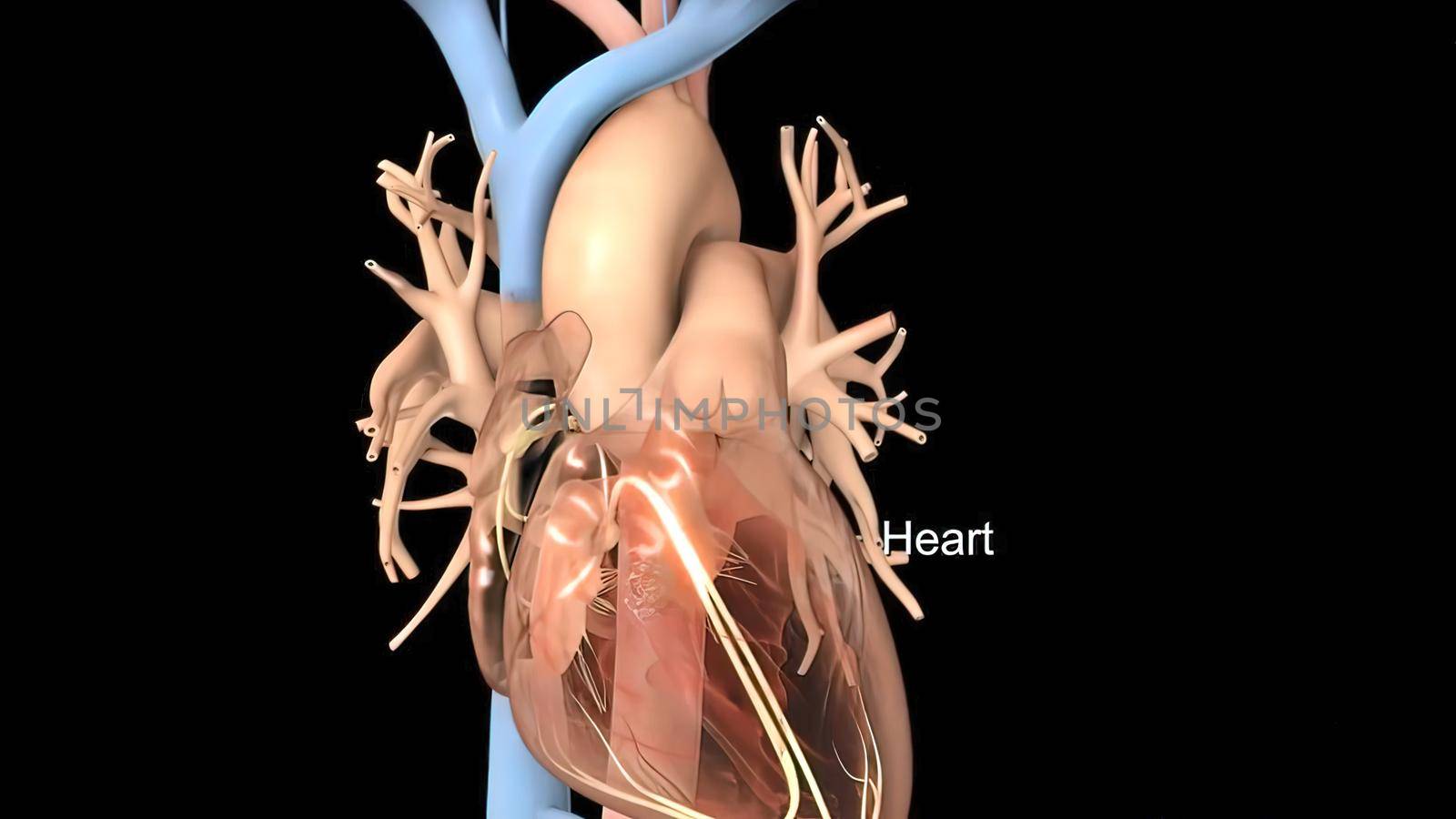
anatomical human heart illustration
Stock PhotoUsername
creativepicResolution
3840x2160pxanatomical human heart illustration
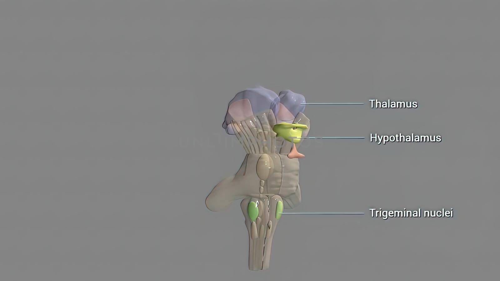
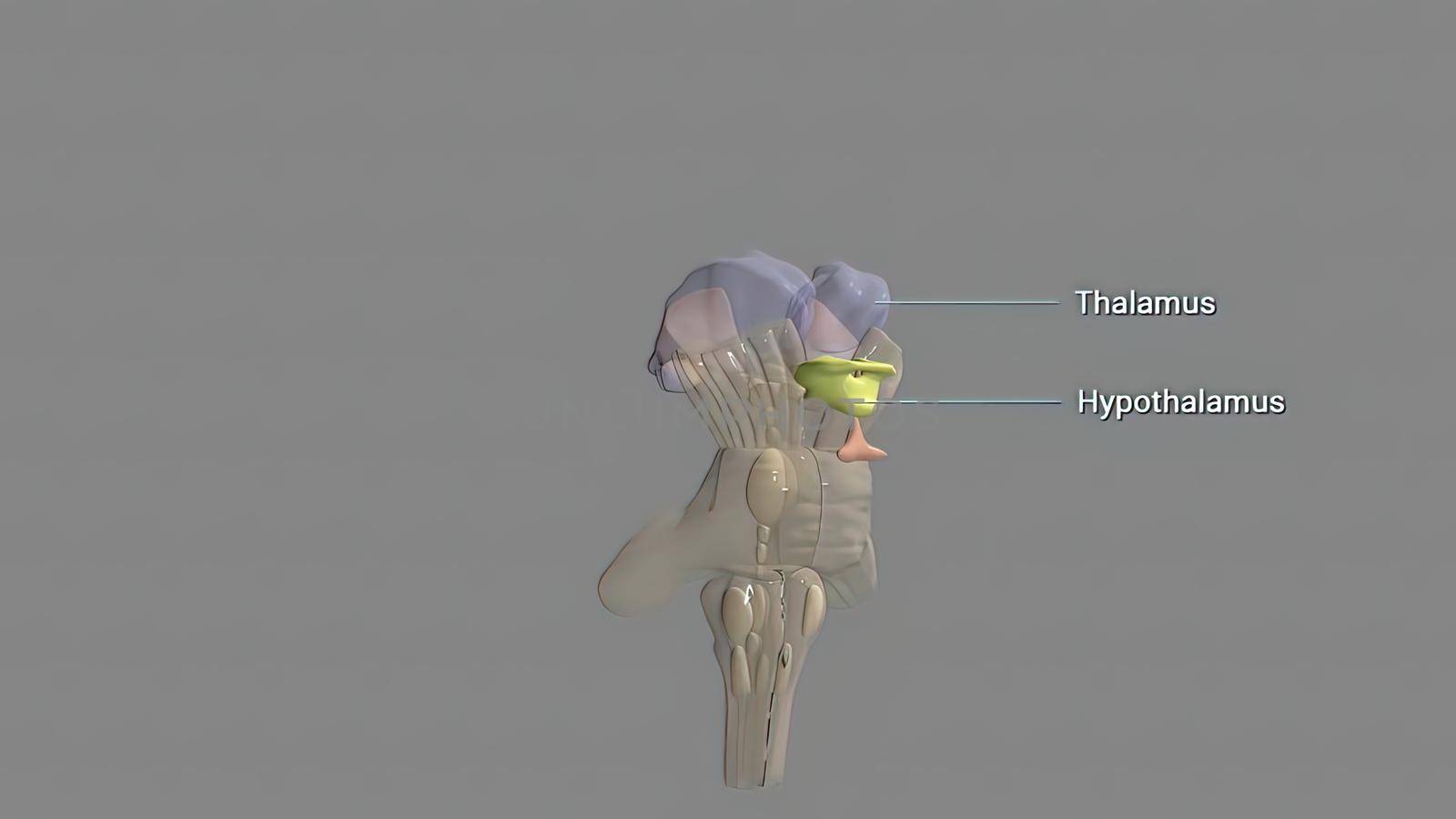
The hypothalamus, highlighted in red, is responsible f
Stock PhotoUsername
creativepicResolution
3840x2160pxThe hypothalamus, highlighted in red, is responsible f
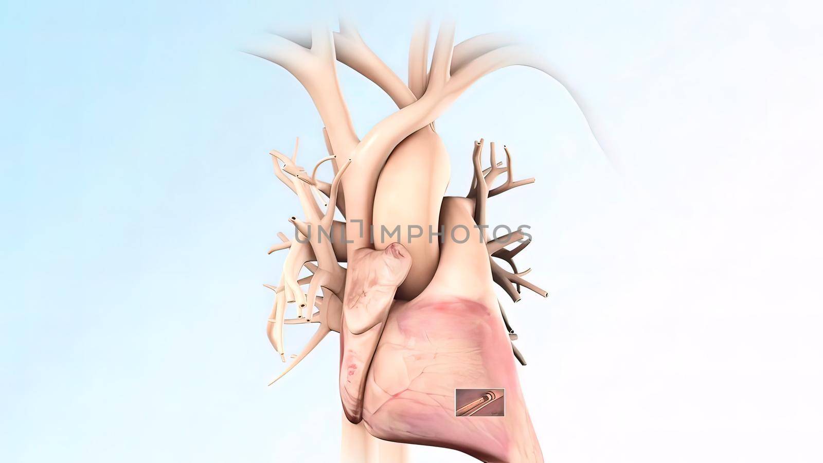
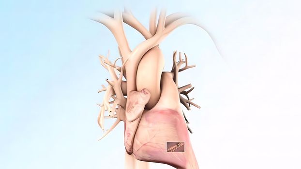
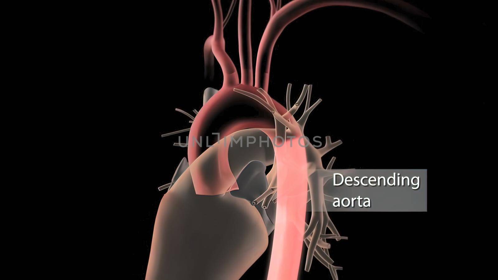
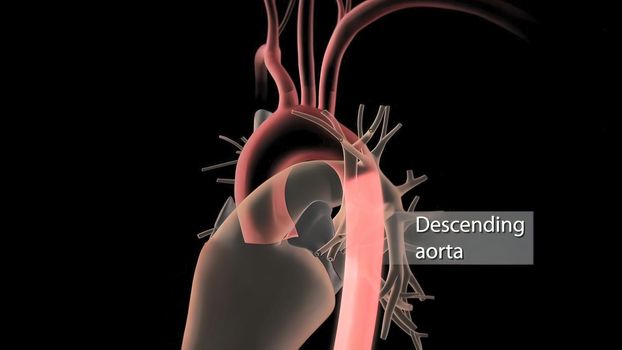
medically accurate illustration of a heart with 3 bypasses
Stock PhotoUsername
creativepicResolution
3840x2160pxmedically accurate illustration of a heart with 3 bypasses
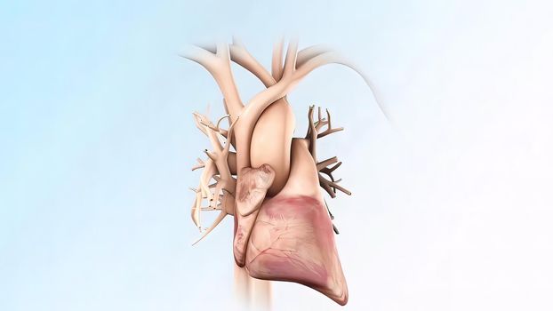
Heart beat. Right side pumps deoxygenated blood and left side oxygenated blood.
Stock PhotoUsername
creativepicResolution
3840x2160pxHeart beat. Right side pumps deoxygenated blood and left side oxygenated blood.

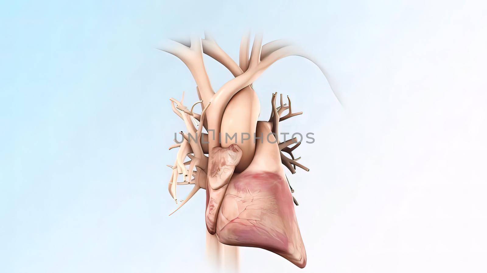
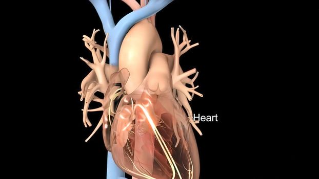
anatomical human heart illustration
Stock PhotoUsername
creativepicResolution
3840x2160pxanatomical human heart illustration

Human Gall Bladder Anatomy With Digestive System For Medical Concept
Stock PhotoUsername
creativepicResolution
3840x2160pxHuman Gall Bladder Anatomy With Digestive System For Medical Concept
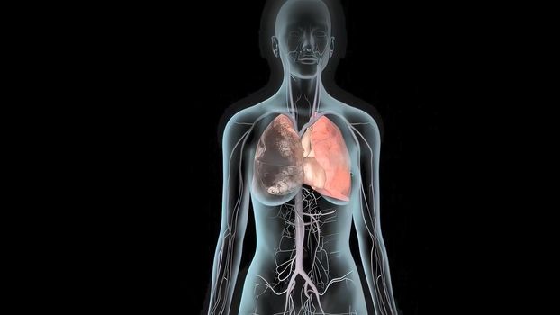
Damaged sick lungs. 3D render illustration for respiratory problems, cancer medical or health problems.
Stock PhotoUsername
creativepicResolution
3840x2160pxDamaged sick lungs. 3D render illustration for respiratory problems, cancer medical or health problems.
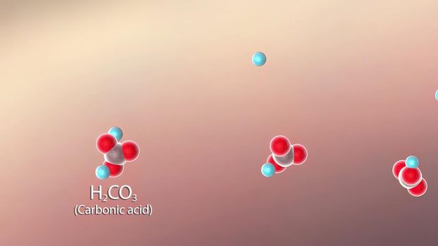
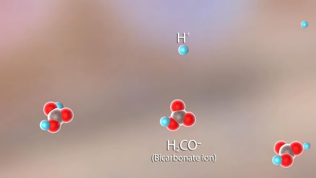
accumulation of carbon dioxide in the blood due to shortness of breath
Stock PhotoUsername
creativepicResolution
3840x2160pxaccumulation of carbon dioxide in the blood due to shortness of breath
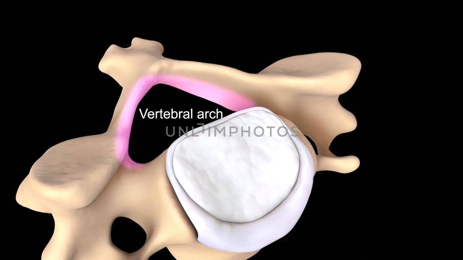
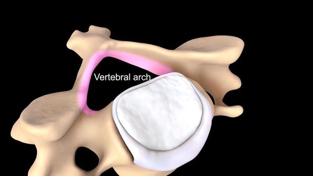
Realistic skeletal human spine and vertebral column or intervertebral discs on a dark background.
Stock PhotoUsername
creativepicResolution
3840x2160pxRealistic skeletal human spine and vertebral column or intervertebral discs on a dark background.

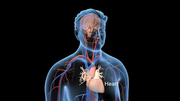
Animated transparent man blood circulation system
Stock PhotoUsername
creativepicResolution
3840x2160pxAnimated transparent man blood circulation system
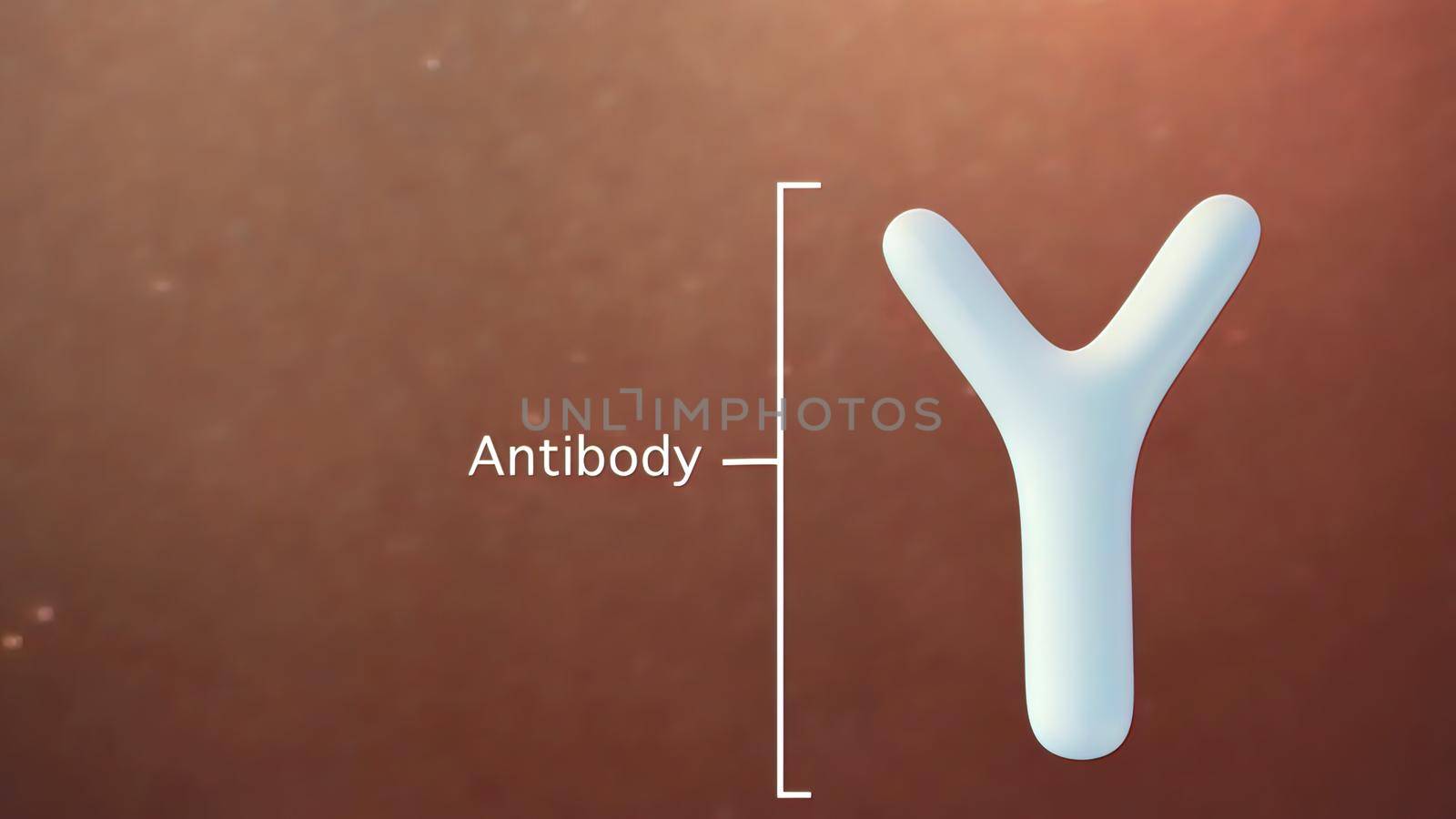
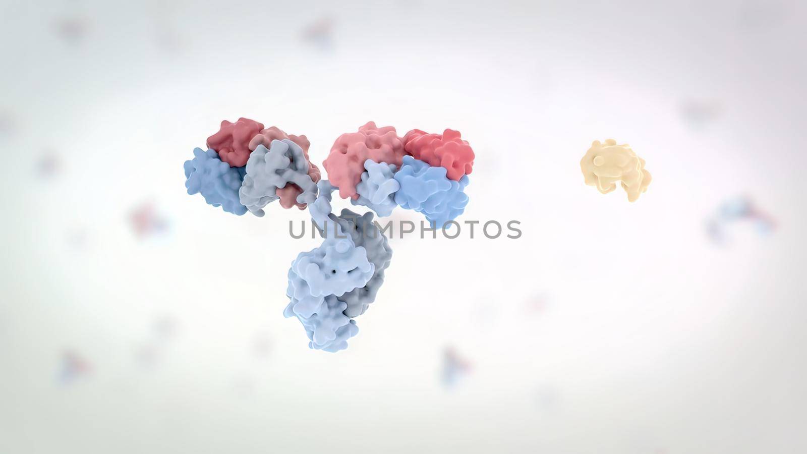
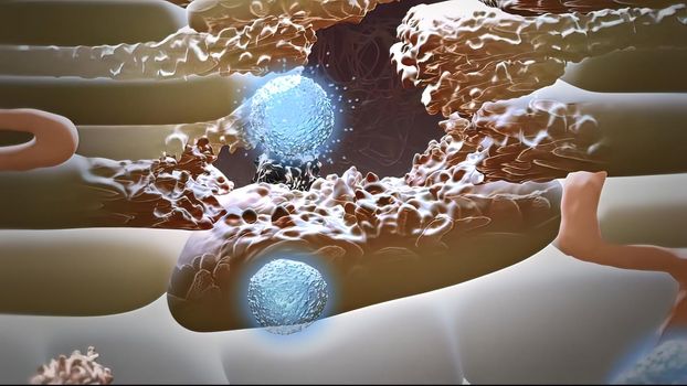
Antibodies that are part of our immune system
Stock PhotoUsername
creativepicResolution
3840x2160pxAntibodies that are part of our immune system

Normal blood pressure 3d medical
Stock PhotoUsername
creativepicResolution
3840x2160pxNormal blood pressure 3d medical
accumulation of carbon dioxide in the blood due to shortness of breath
Stock PhotoUsername
creativepicResolution
3840x2160pxaccumulation of carbon dioxide in the blood due to shortness of breath

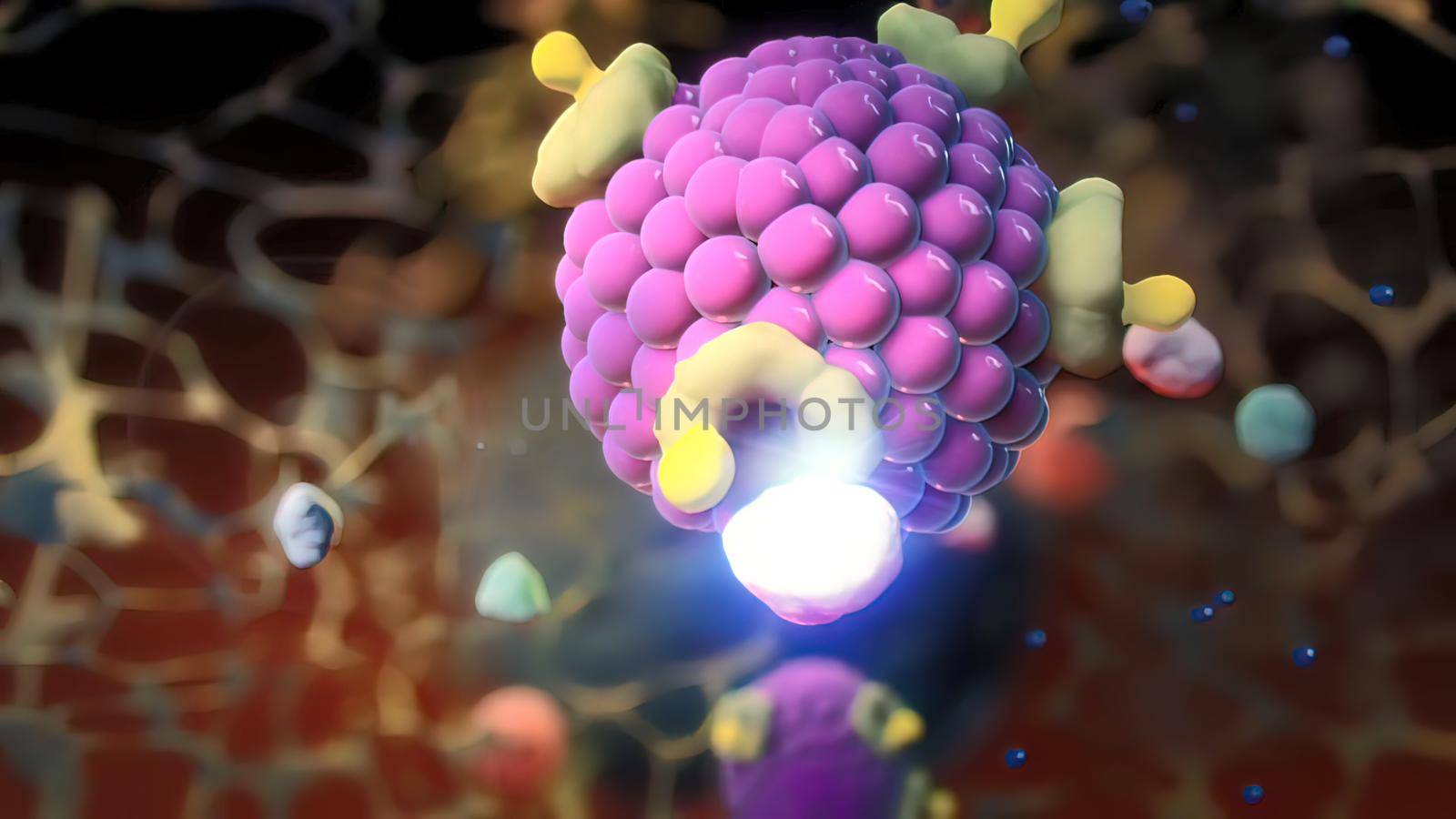
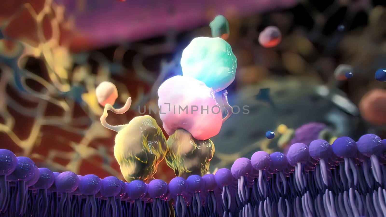
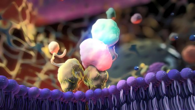
Receptors, transport of monoclonal antibodies across the blood-brain barrier 3D medical illustration
Stock PhotoUsername
creativepicResolution
3840x2160pxReceptors, transport of monoclonal antibodies across the blood-brain barrier 3D medical illustration
Sinoatrial node and cardiovascular system
Stock PhotoUsername
creativepicResolution
3840x2160pxSinoatrial node and cardiovascular system

blood clot in the lung
Stock PhotoUsername
creativepicResolution
3840x2160pxblood clot in the lung
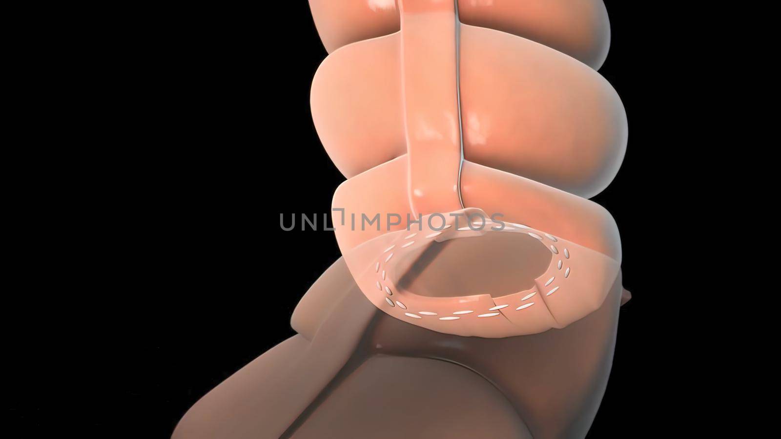
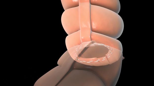
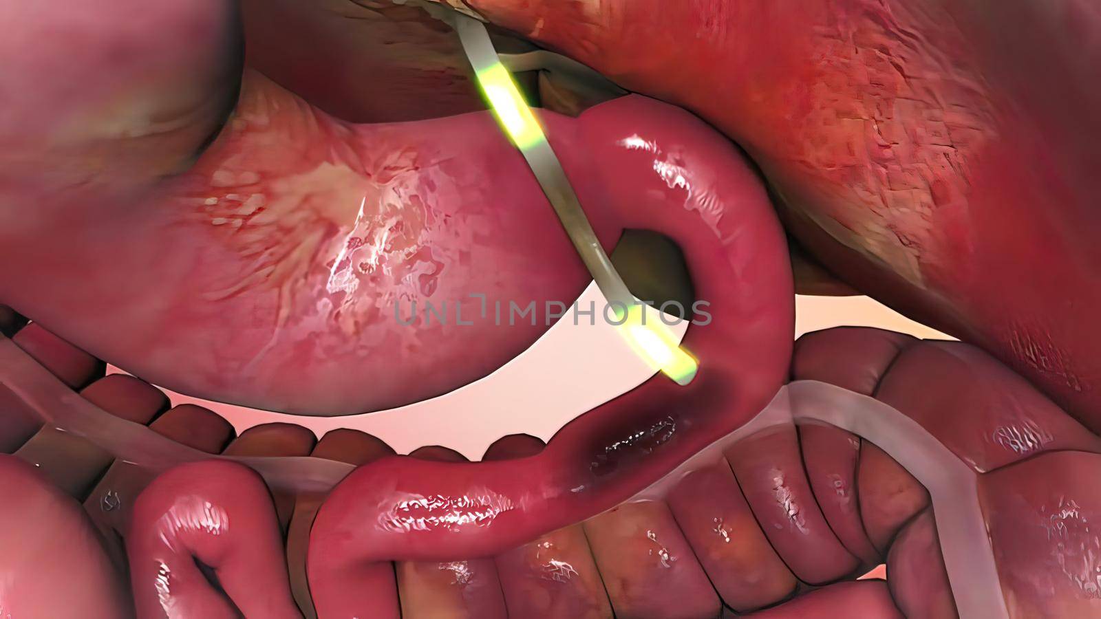
surgery to remove any part of the intestines, bowel resection
Stock PhotoUsername
creativepicResolution
3840x2160pxsurgery to remove any part of the intestines, bowel resection
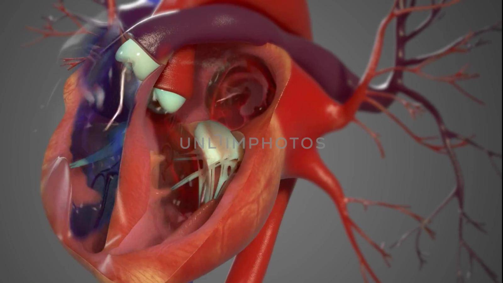
Human Circulatory System Heart Beat Anatomy 3D Render Concept.
Stock PhotoUsername
creativepicResolution
3840x2160pxHuman Circulatory System Heart Beat Anatomy 3D Render Concept.
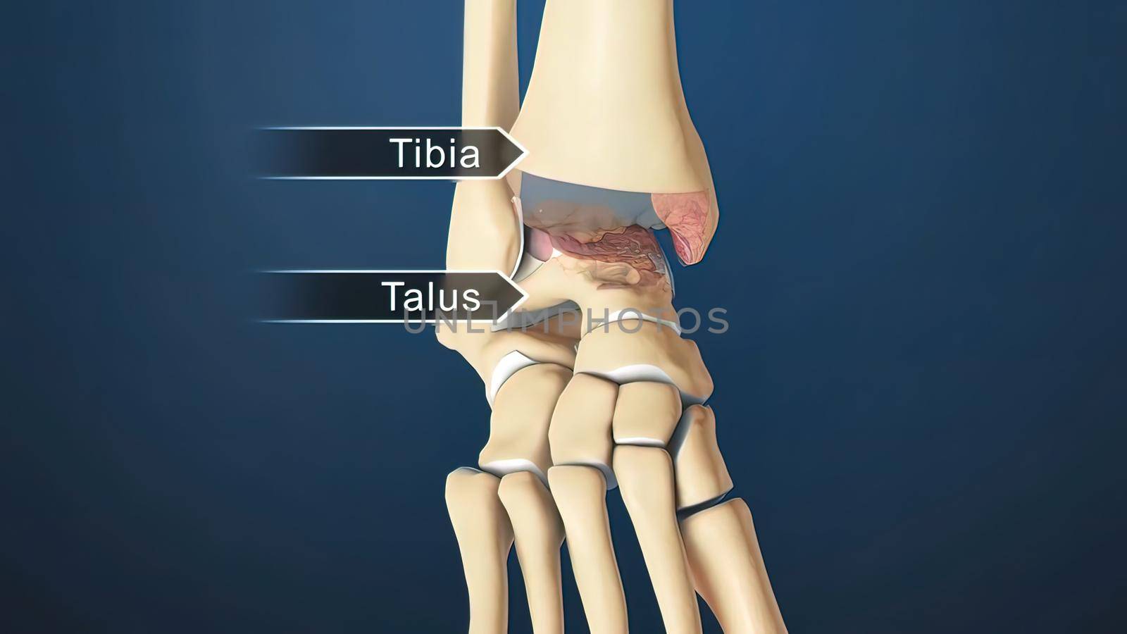
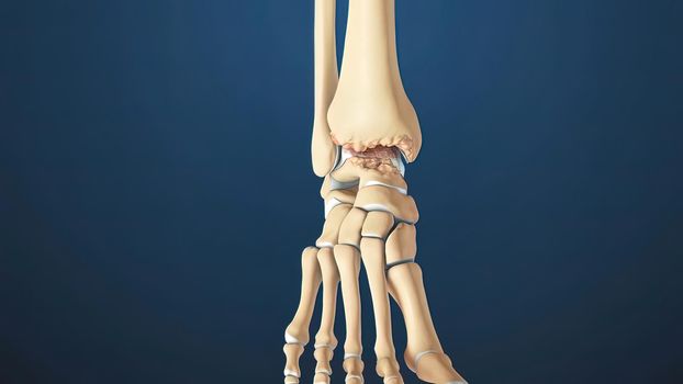
Ankle Joint Anatomy and articular cartilage
Stock PhotoUsername
creativepicResolution
3840x2160pxAnkle Joint Anatomy and articular cartilage
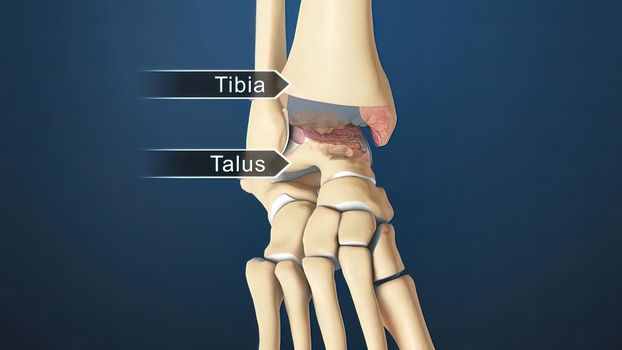

Ankle Joint Anatomy and articular cartilage
Stock PhotoUsername
creativepicResolution
3840x2160pxAnkle Joint Anatomy and articular cartilage
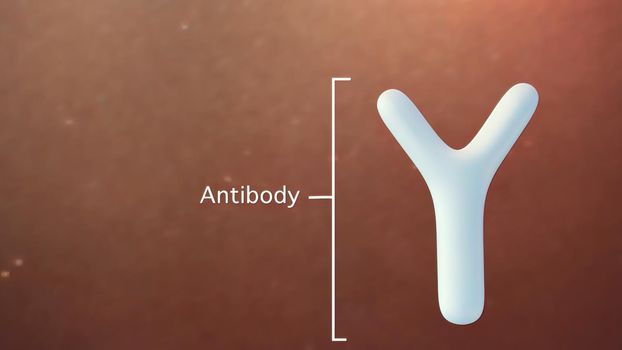
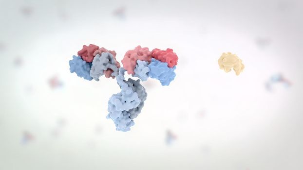
Antibodies are proteins produced by the immune system to fight infections.
Stock PhotoUsername
creativepicResolution
3840x2160pxAntibodies are proteins produced by the immune system to fight infections.
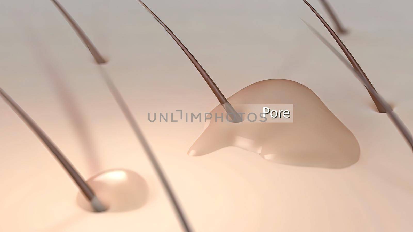
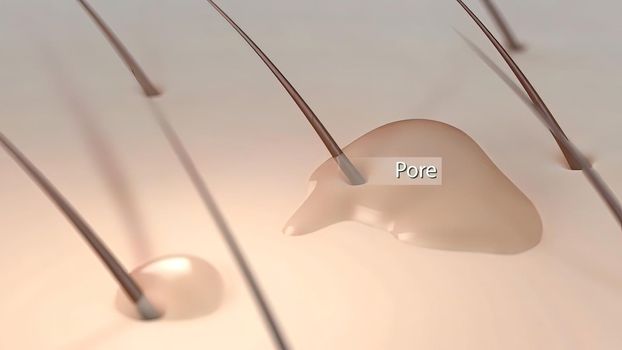
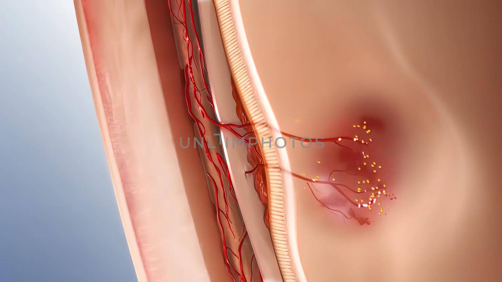
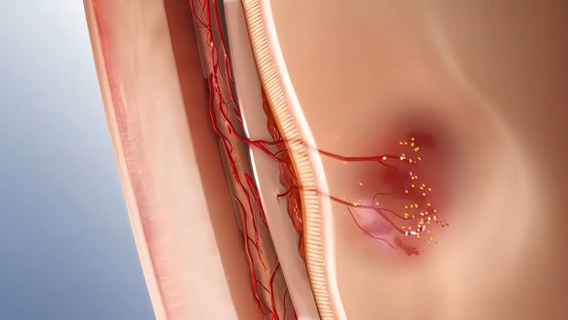
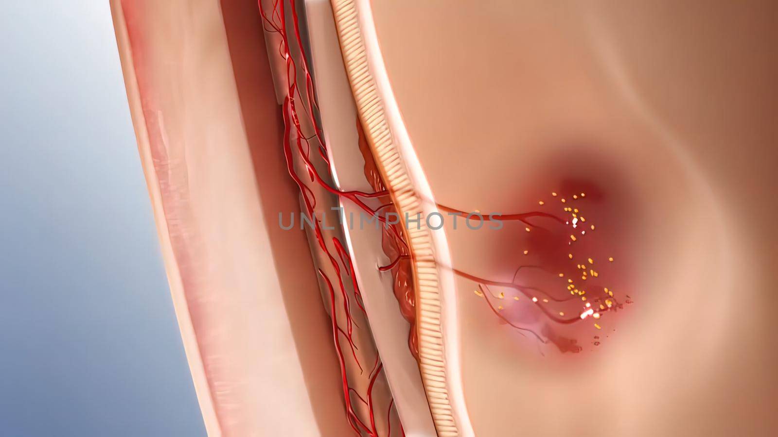
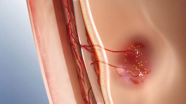
inflammatory acne occurs when bacteria infect clogged or closed pores
Stock PhotoUsername
creativepicResolution
3840x2160pxinflammatory acne occurs when bacteria infect clogged or closed pores
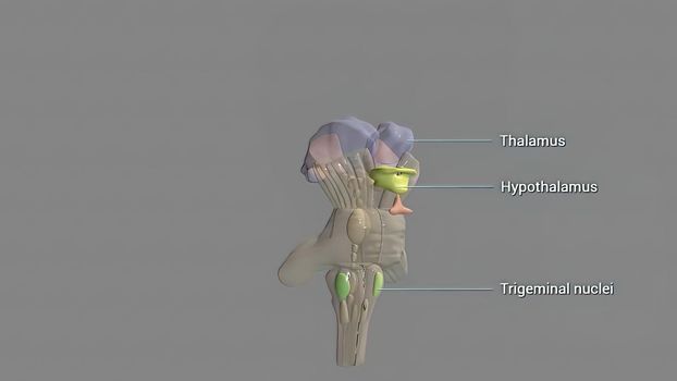
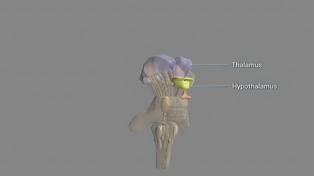
The hypothalamus, highlighted in red, is responsible f
Stock PhotoUsername
creativepicResolution
3840x2160pxThe hypothalamus, highlighted in red, is responsible f

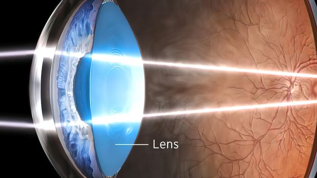
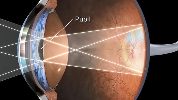
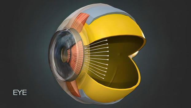
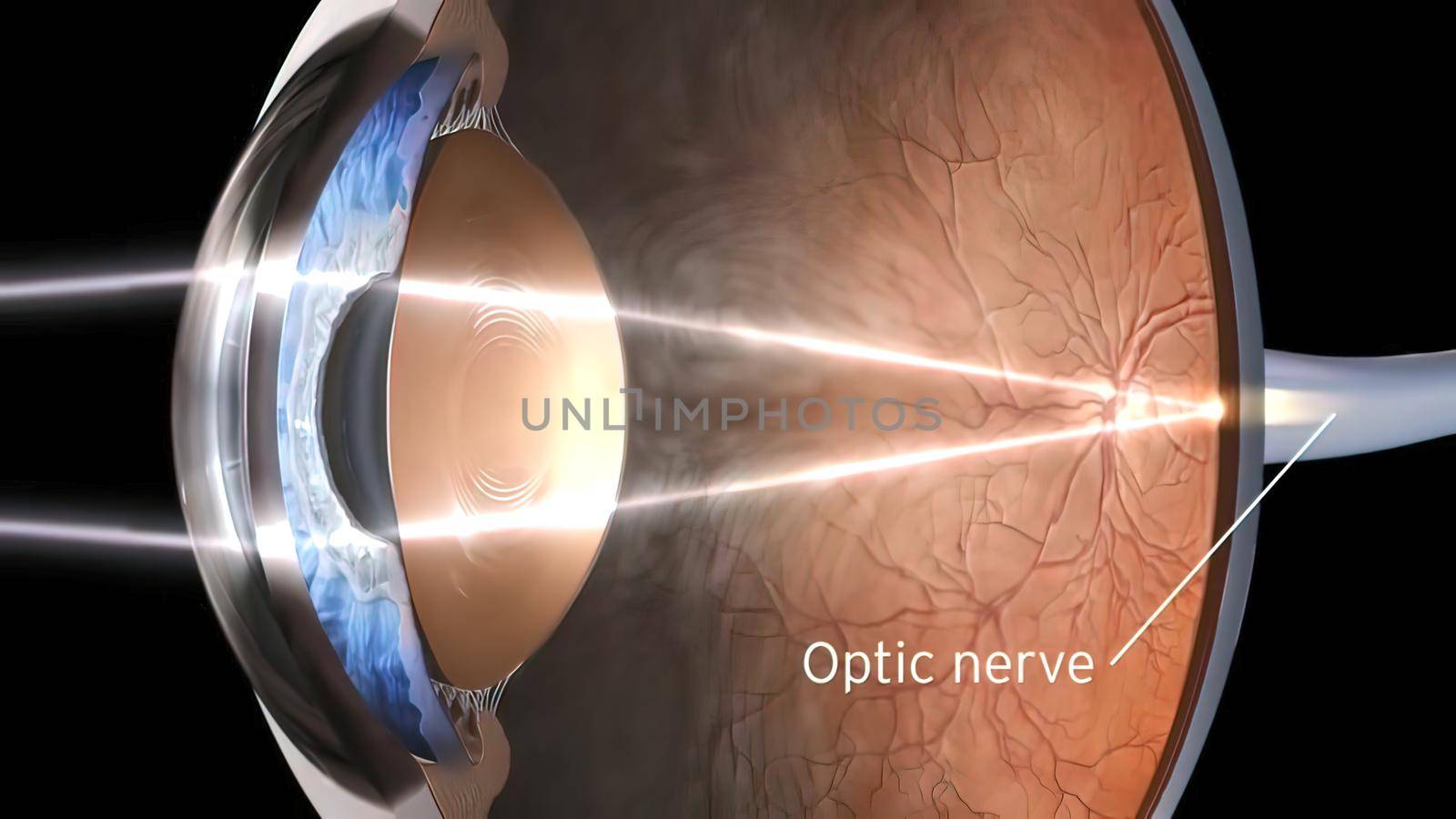
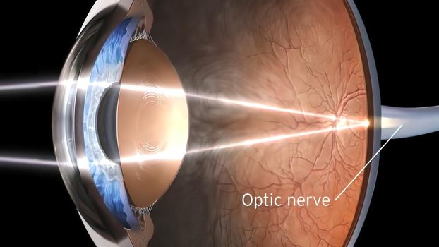
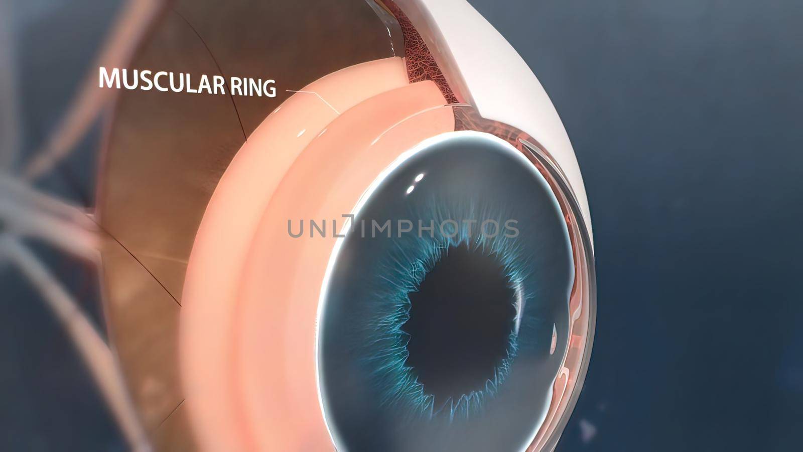
Eye Anatomy - Internal Structure, Medically Accurate
Stock PhotoUsername
creativepicResolution
7680x4320pxEye Anatomy - Internal Structure, Medically Accurate
human internal organs.Role of liver on organs
Stock PhotoUsername
creativepicResolution
7680x4320pxhuman internal organs.Role of liver on organs


Kidney with growing stones
Stock PhotoUsername
creativepicResolution
7680x4320pxKidney with growing stones

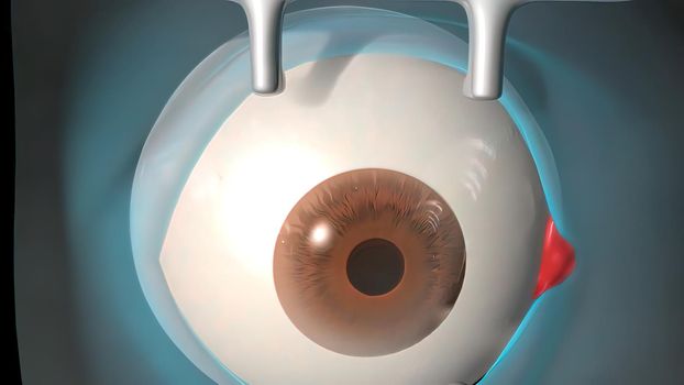
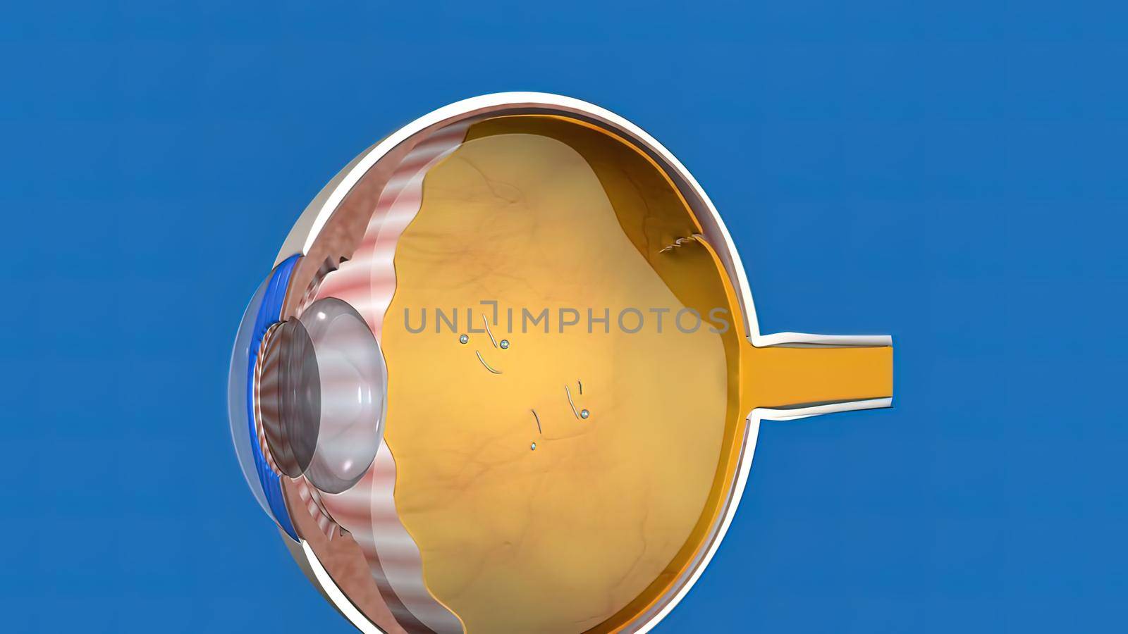
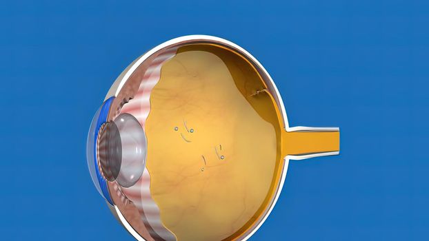
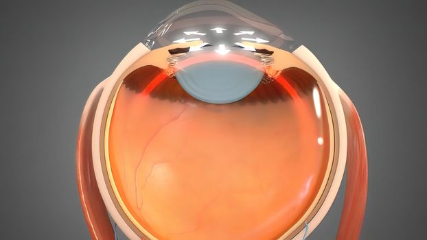
Cataract surgery application view Surgical operations on the human eye
Stock PhotoUsername
creativepicResolution
7680x4320pxCataract surgery application view Surgical operations on the human eye
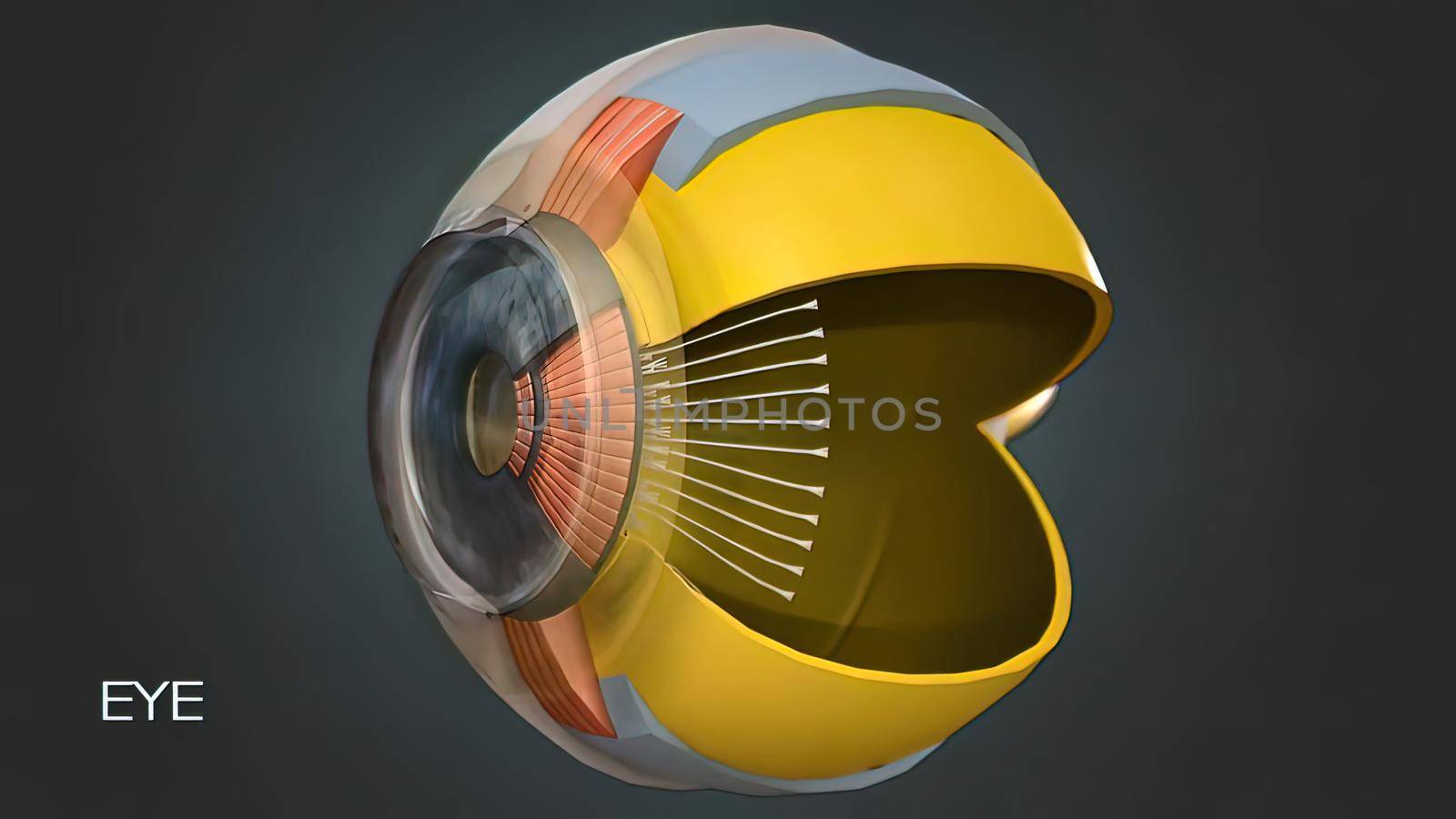
Eye Anatomy - Internal Structure, Medically Accurate
Stock PhotoUsername
creativepicResolution
7680x4320pxEye Anatomy - Internal Structure, Medically Accurate

Epigenetic Regulation of Excitatory Amino Acid Transporter in Neurological Disorders
Stock PhotoUsername
creativepicResolution
7680x4320pxEpigenetic Regulation of Excitatory Amino Acid Transporter in Neurological Disorders

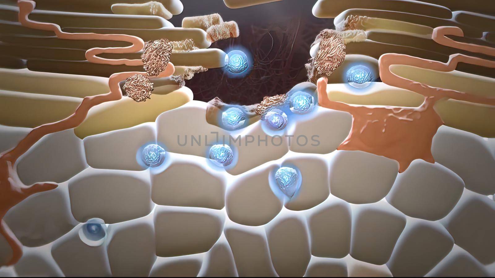
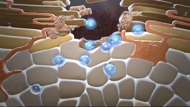
Ion Channels in Epithelial Cells
Stock PhotoUsername
creativepicResolution
7680x4320pxIon Channels in Epithelial Cells

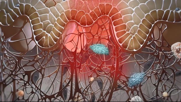
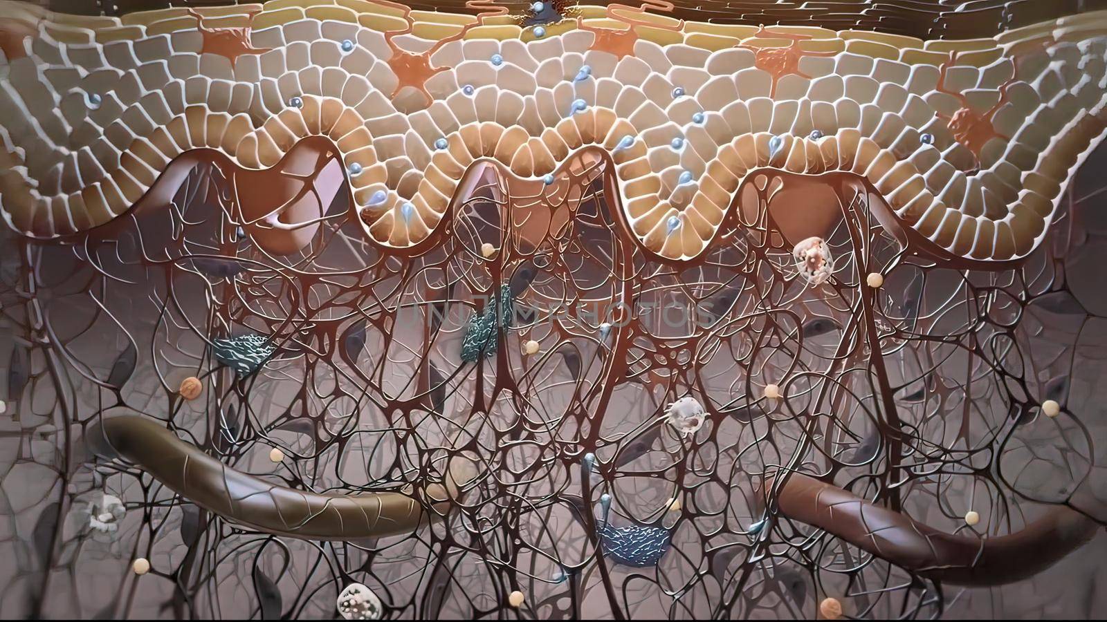
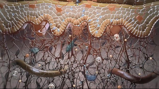
In the layer adjacent to the dermis, the keratinocytes are divided and thrown into the upper layers.
Stock PhotoUsername
creativepicResolution
7680x4320pxIn the layer adjacent to the dermis, the keratinocytes are divided and thrown into the upper layers.

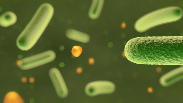
Microscopic visualization of bacteria .
Stock PhotoUsername
creativepicResolution
7680x4320pxMicroscopic visualization of bacteria .

In the layer adjacent to the dermis, the keratinocytes are divided and thrown into the upper layers.
Stock PhotoUsername
creativepicResolution
7680x4320pxIn the layer adjacent to the dermis, the keratinocytes are divided and thrown into the upper layers.
Ion Channels in Epithelial Cells
Stock PhotoUsername
creativepicResolution
7680x4320pxIon Channels in Epithelial Cells
In the layer adjacent to the dermis, the keratinocytes are divided and thrown into the upper layers.
Stock PhotoUsername
creativepicResolution
7680x4320pxIn the layer adjacent to the dermis, the keratinocytes are divided and thrown into the upper layers.

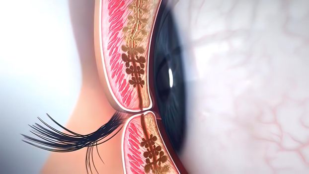
Closure of the eyelids, side view
Stock PhotoUsername
creativepicResolution
7680x4320pxClosure of the eyelids, side view
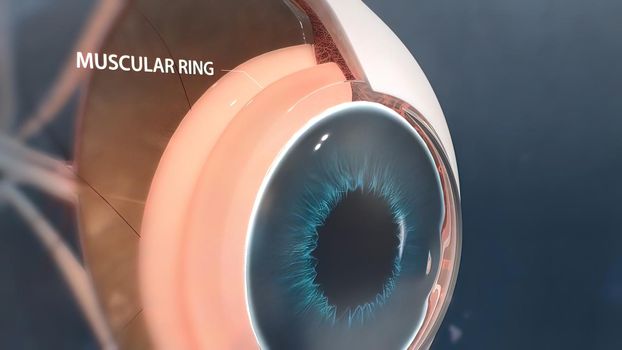
Eye Anatomy - Internal Structure, Medically Accurate
Stock PhotoUsername
creativepicResolution
7680x4320pxEye Anatomy - Internal Structure, Medically Accurate
3D illustration medical animated eye anatomy, eye stain formation
Stock PhotoUsername
creativepicResolution
7680x4320px3D illustration medical animated eye anatomy, eye stain formation
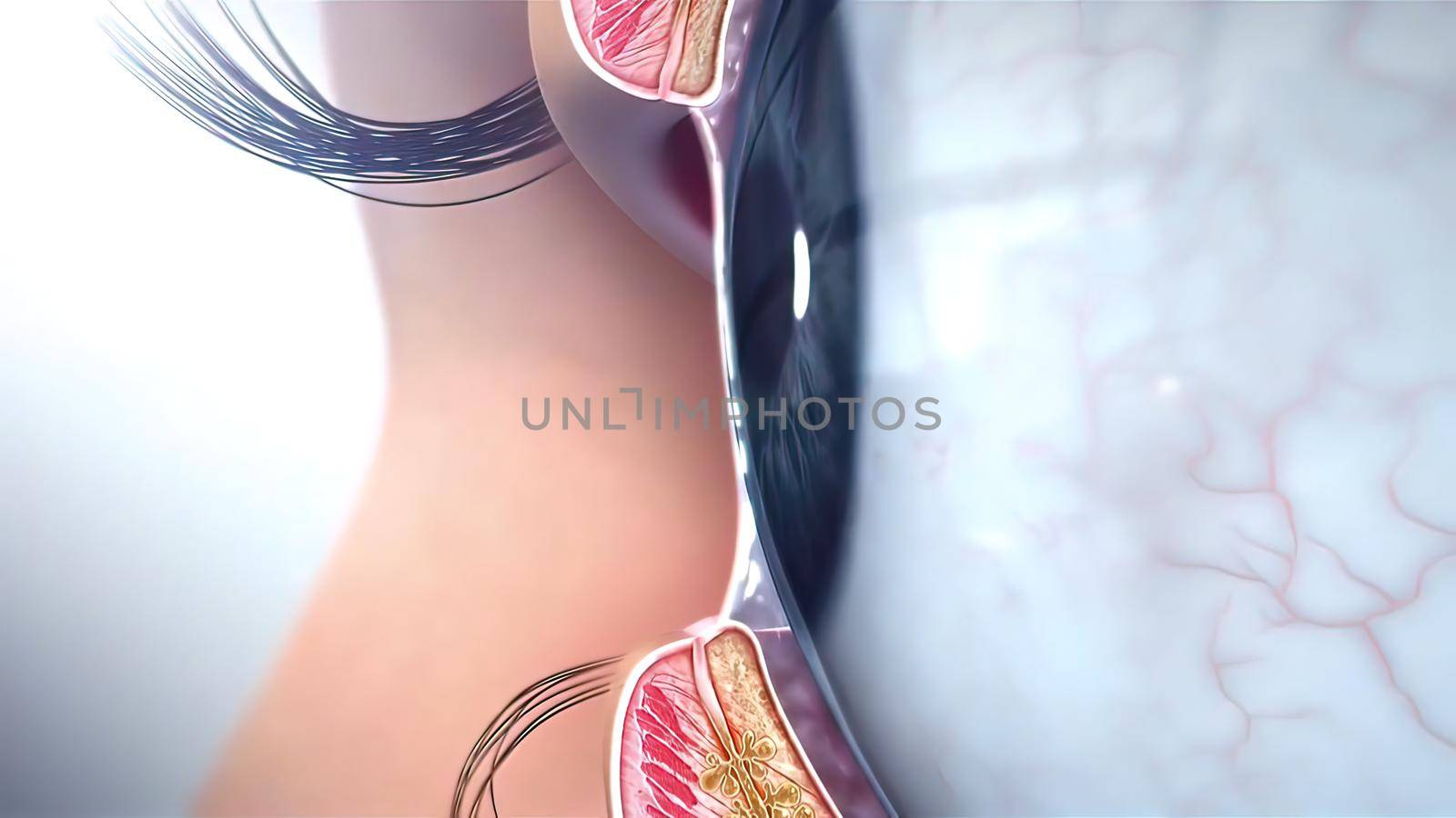
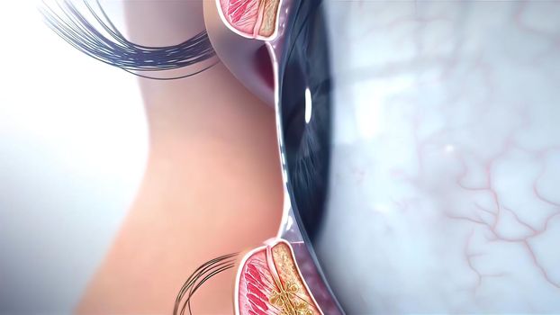
Laser eye surgery, Eyes anatomy
Stock PhotoUsername
creativepicResolution
7680x4320pxLaser eye surgery, Eyes anatomy

In the layer adjacent to the dermis, the keratinocytes are divided and thrown into the upper layers.
Stock PhotoUsername
creativepicResolution
7680x4320pxIn the layer adjacent to the dermis, the keratinocytes are divided and thrown into the upper layers.
Glaucoma eye disease 3D illustration medical
Stock PhotoUsername
creativepicResolution
7680x4320pxGlaucoma eye disease 3D illustration medical
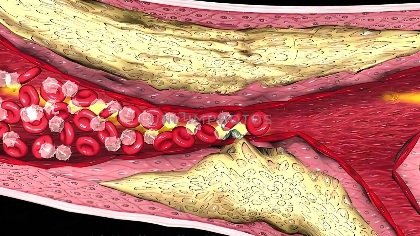
Coronary atherosclerosis, light micrograph showing cholesterol-containing plaque
Stock PhotoUsername
creativepicResolution
3840x2160pxCoronary atherosclerosis, light micrograph showing cholesterol-containing plaque


Green Eye of Child Looking at Camera Close Up. Macro Shot Opening
Stock PhotoUsername
creativepicResolution
7680x4320pxGreen Eye of Child Looking at Camera Close Up. Macro Shot Opening
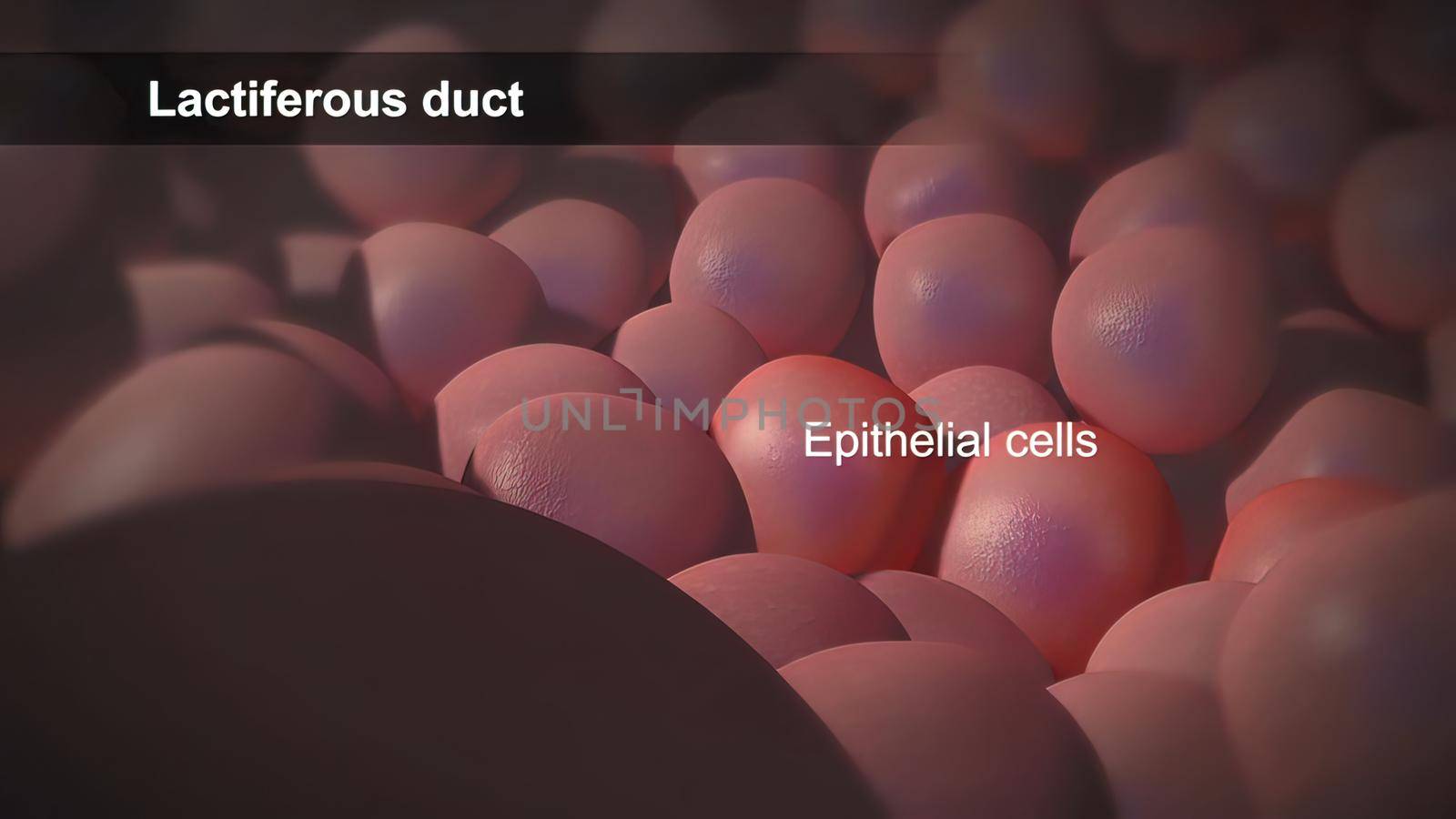
Epithelial cells are a type of cell that lines the surfaces of your body.
Stock PhotoUsername
creativepicResolution
7680x4320pxEpithelial cells are a type of cell that lines the surfaces of your body.

Glaucoma eye disease 3D illustration medical
Stock PhotoUsername
creativepicResolution
7680x4320pxGlaucoma eye disease 3D illustration medical

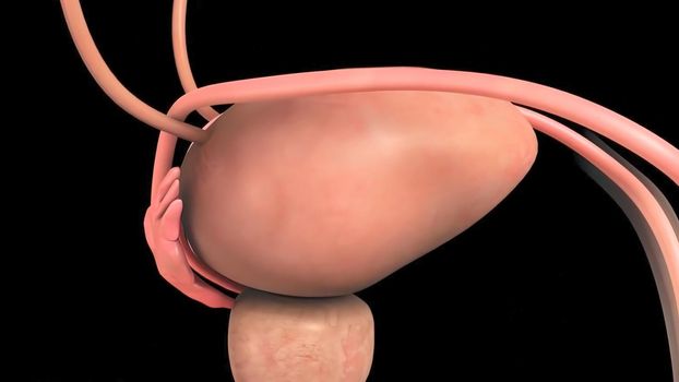
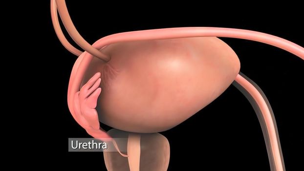
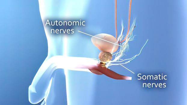
Male reproductive system 3D illustration
Stock PhotoUsername
creativepicResolution
7680x4320pxMale reproductive system 3D illustration
Eye Anatomy - Internal Structure, Medically Accurate
Stock PhotoUsername
creativepicResolution
7680x4320pxEye Anatomy - Internal Structure, Medically Accurate

Eye Anatomy - Internal Structure, Medically Accurate
Stock PhotoUsername
creativepicResolution
7680x4320pxEye Anatomy - Internal Structure, Medically Accurate