- Filter By:
-
-
Stock photos and images of username:stockdevil

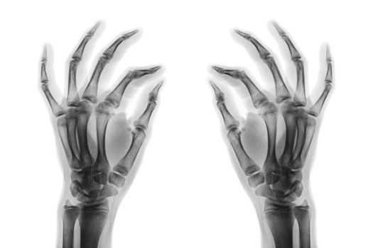
Fracture at 3rd and 4th metacarpal bone . Film x-ray of adult hands . Oblique view
Stock PhotoUsername
stockdevilResolution
5184x3456pxFracture at 3rd and 4th metacarpal bone . Film x-ray of adult hands . Oblique view

Fracture at 3rd and 4th metacarpal bone . Film x-ray of adult hands . Oblique view
Stock PhotoUsername
stockdevilResolution
5184x3456pxFracture at 3rd and 4th metacarpal bone . Film x-ray of adult hands . Oblique view

Fracture at 3rd and 4th metacarpal bone . Film x-ray of adult hands . Oblique view
Stock PhotoUsername
stockdevilResolution
5184x3456pxFracture at 3rd and 4th metacarpal bone . Film x-ray of adult hands . Oblique view

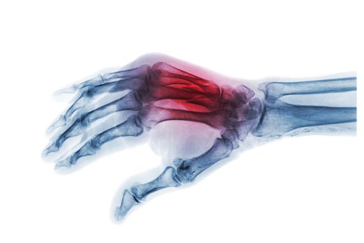
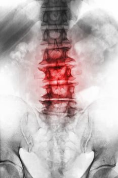
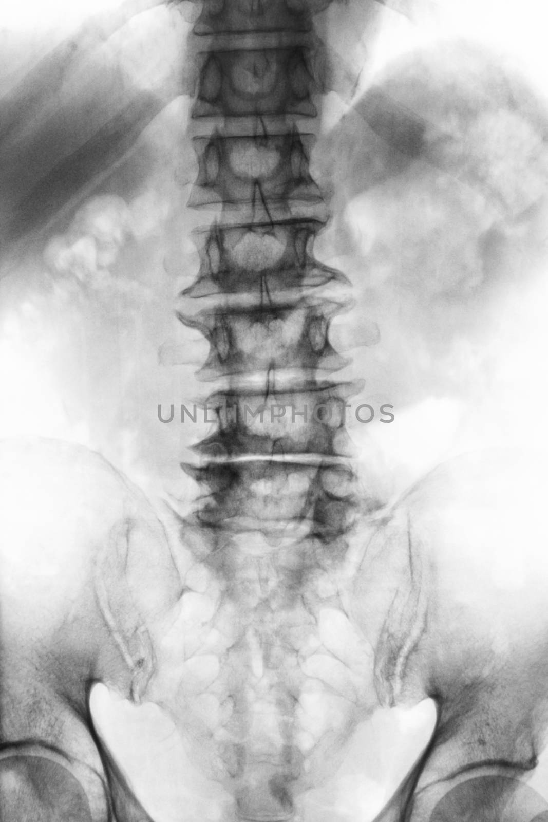
Spondylosis . film x-ray lumbosacral spine of old aged patient show osteophyte , collapse spine from degenerative process . Front view
Stock PhotoUsername
stockdevilResolution
3421x5131pxSpondylosis . film x-ray lumbosacral spine of old aged patient show osteophyte , collapse spine from degenerative process . Front view
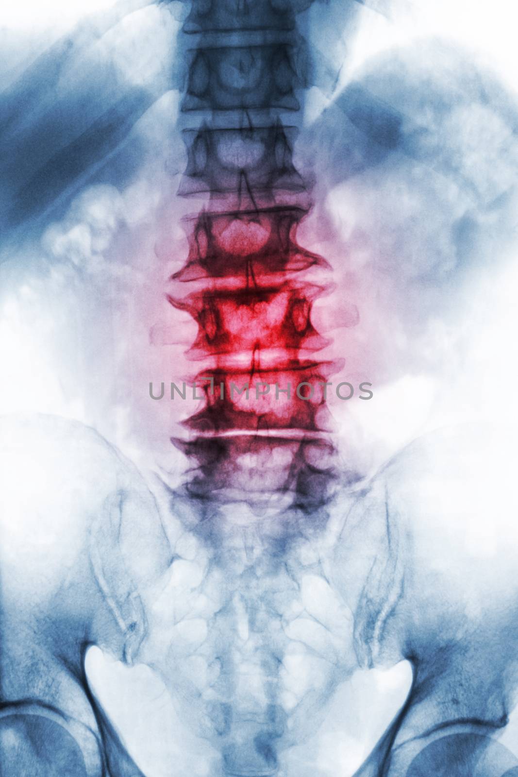
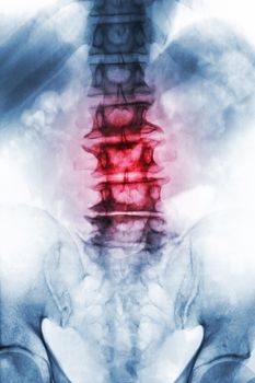
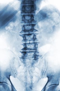
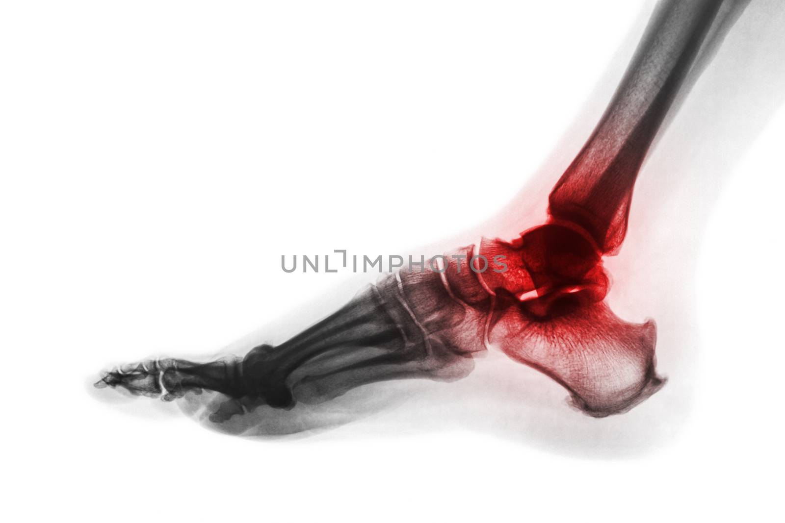
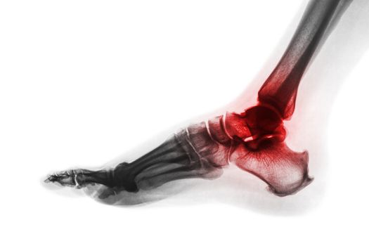
Arthritis of ankle . X-ray of foot . Lateral view . Invert color style . Gout or Rheumatoid concept
Stock PhotoUsername
stockdevilResolution
5184x3456pxArthritis of ankle . X-ray of foot . Lateral view . Invert color style . Gout or Rheumatoid concept

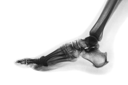
X-ray normal human foot . Lateral view . Invert color style
Stock PhotoUsername
stockdevilResolution
5184x3456pxX-ray normal human foot . Lateral view . Invert color style

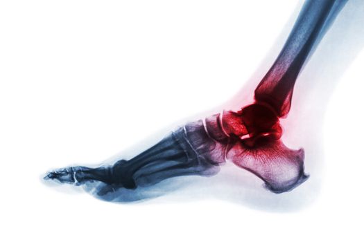
Arthritis of ankle . X-ray of foot . Lateral view . Invert color style . Gout or Rheumatoid concept
Stock PhotoUsername
stockdevilResolution
5184x3456pxArthritis of ankle . X-ray of foot . Lateral view . Invert color style . Gout or Rheumatoid concept
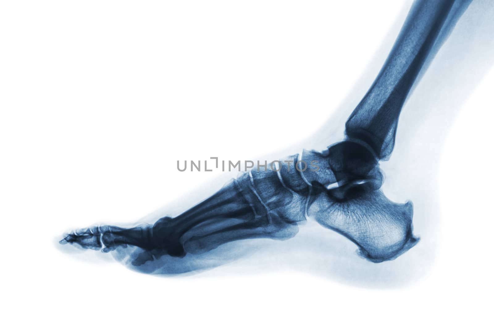
X-ray normal human foot . Lateral view . Invert color style
Stock PhotoUsername
stockdevilResolution
5184x3456pxX-ray normal human foot . Lateral view . Invert color style

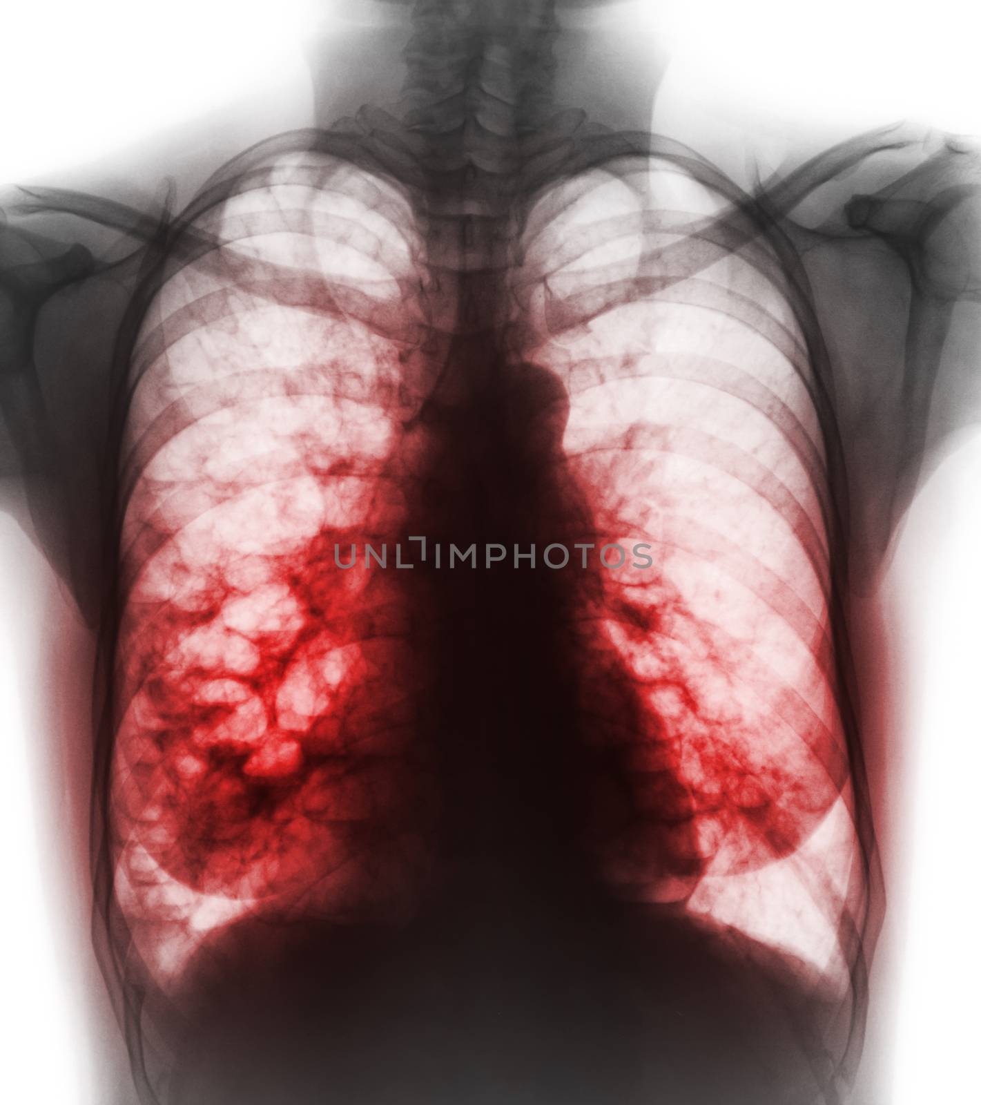
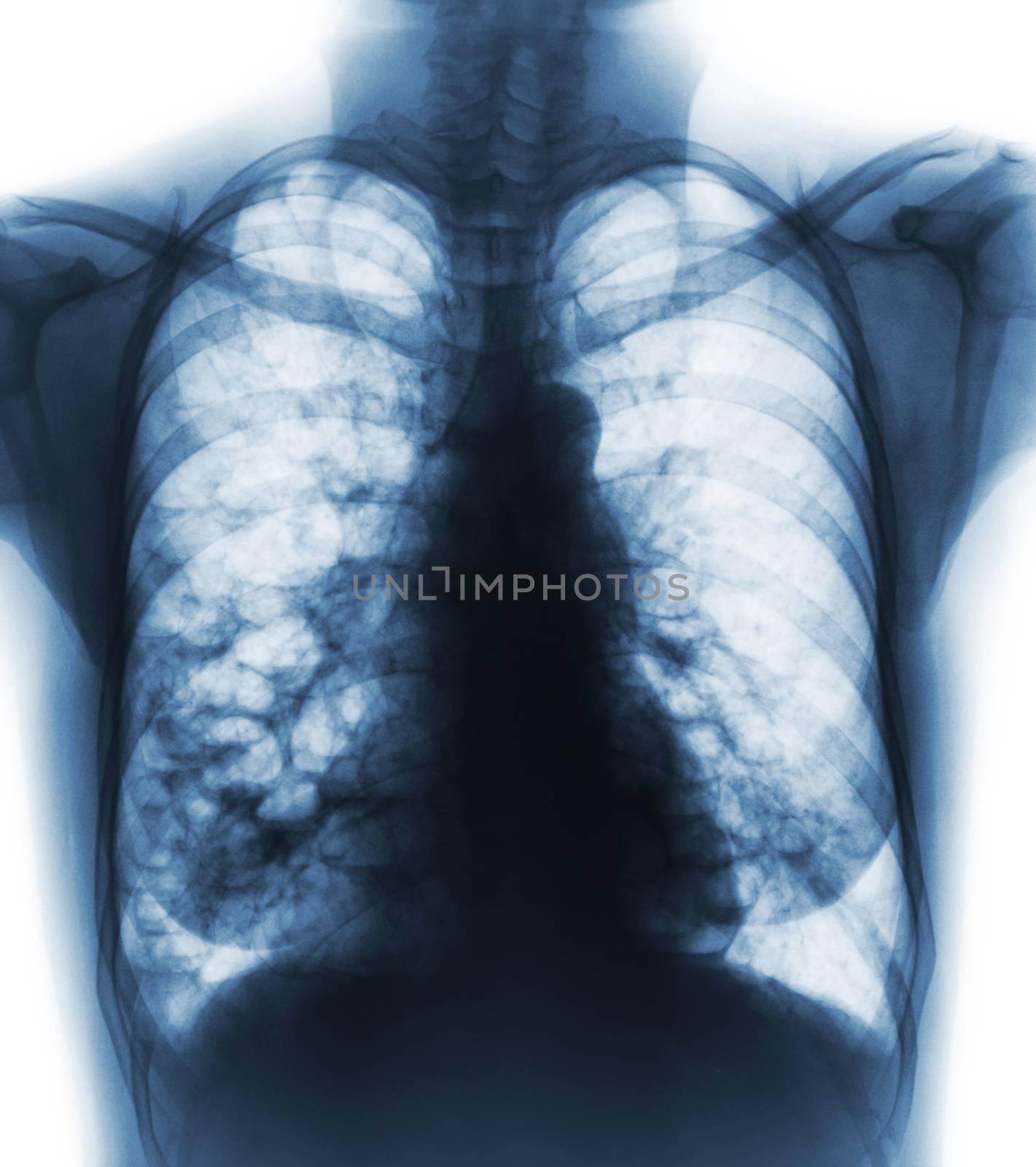
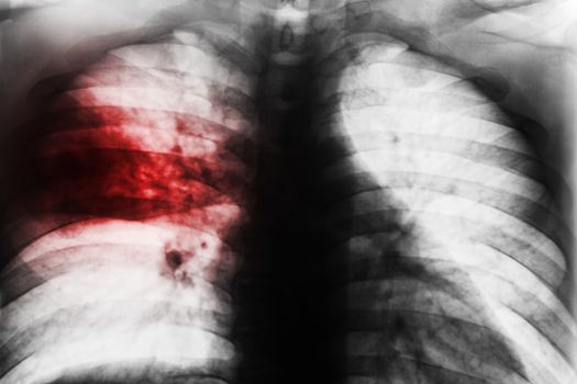
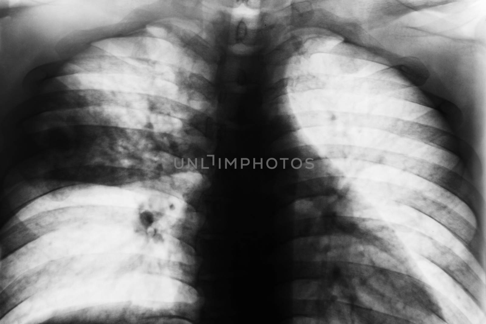
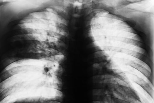
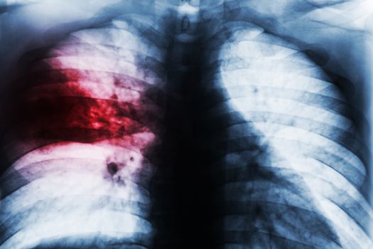
Bronchiectasis . X-ray chest show multiple lung bleb and cyst due to chronic infection . Front view
Stock PhotoUsername
stockdevilResolution
3297x3713pxBronchiectasis . X-ray chest show multiple lung bleb and cyst due to chronic infection . Front view

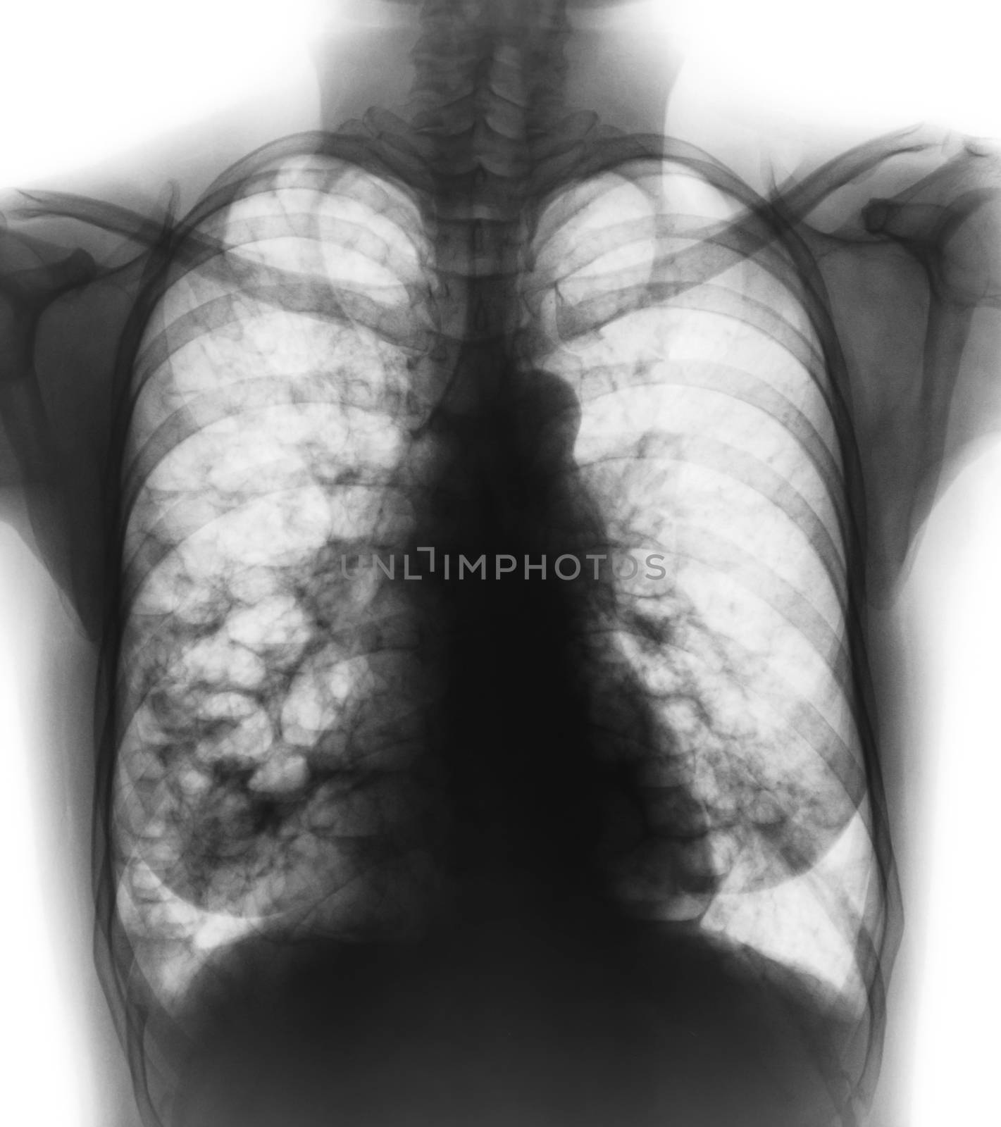
Bronchiectasis . X-ray chest show multiple lung bleb and cyst due to chronic infection . Front view
Stock PhotoUsername
stockdevilResolution
3297x3713pxBronchiectasis . X-ray chest show multiple lung bleb and cyst due to chronic infection . Front view


Bronchiectasis . X-ray chest show multiple lung bleb and cyst due to chronic infection . Front view
Stock PhotoUsername
stockdevilResolution
3297x3713pxBronchiectasis . X-ray chest show multiple lung bleb and cyst due to chronic infection . Front view
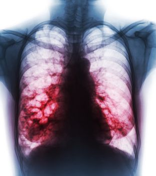
Bronchiectasis . X-ray chest show multiple lung bleb and cyst due to chronic infection . Front view
Stock PhotoUsername
stockdevilResolution
3297x3713pxBronchiectasis . X-ray chest show multiple lung bleb and cyst due to chronic infection . Front view

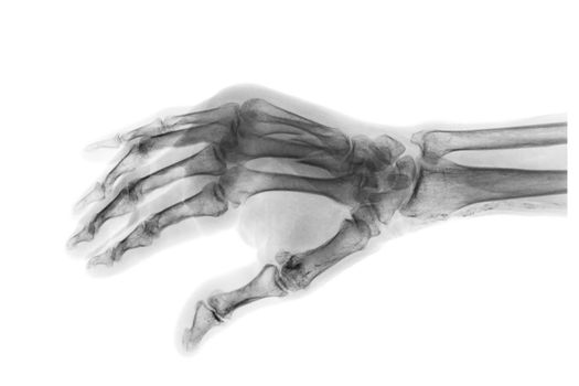
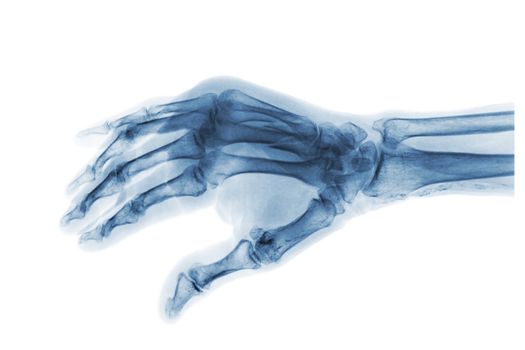
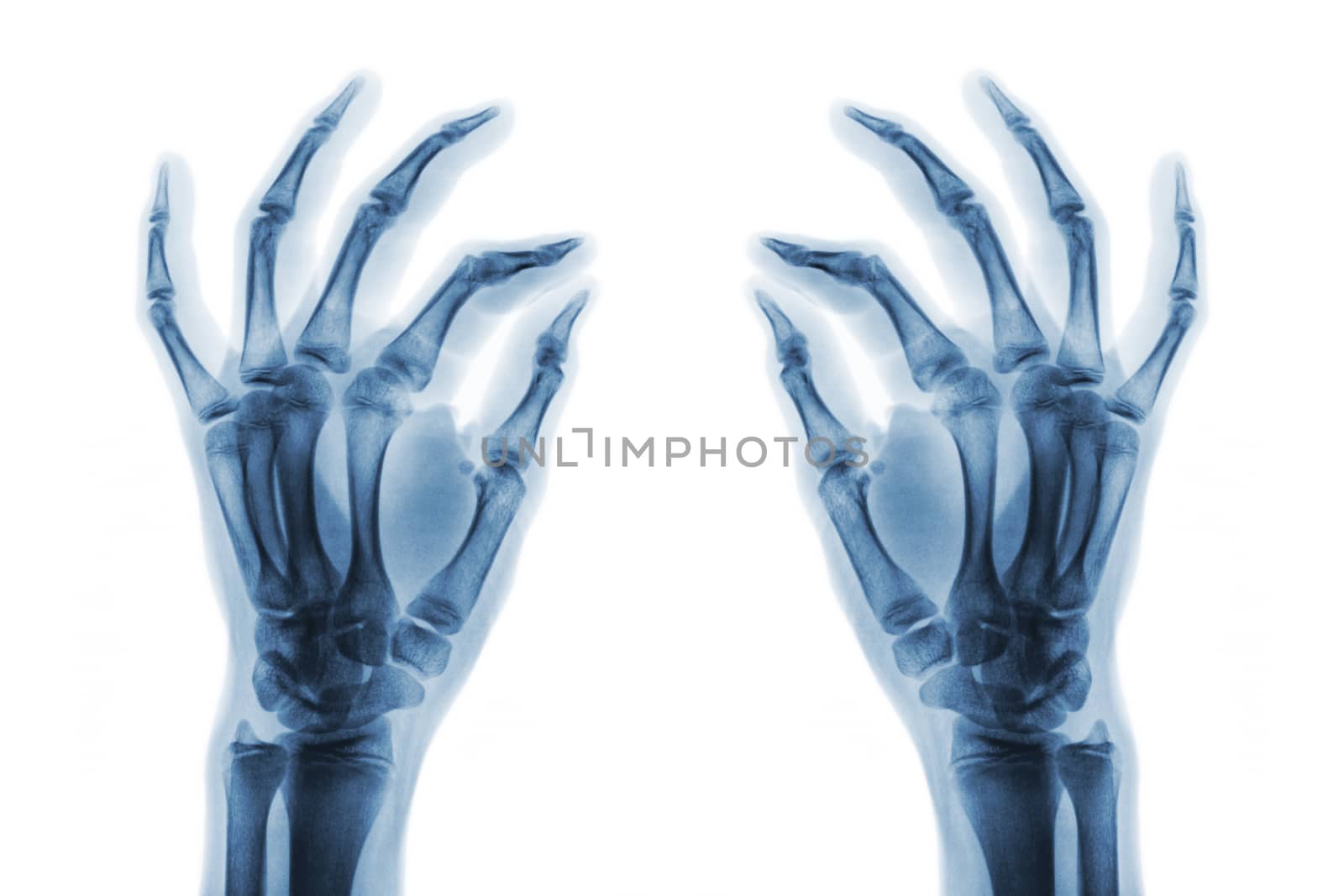
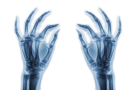
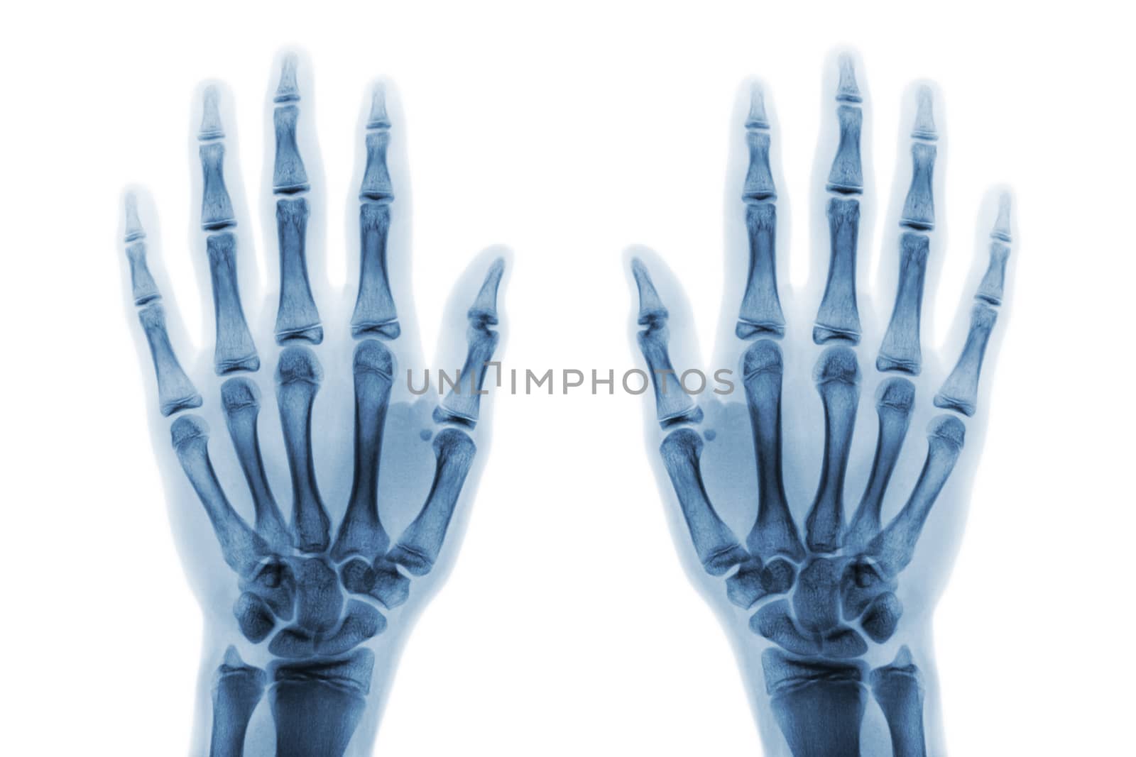
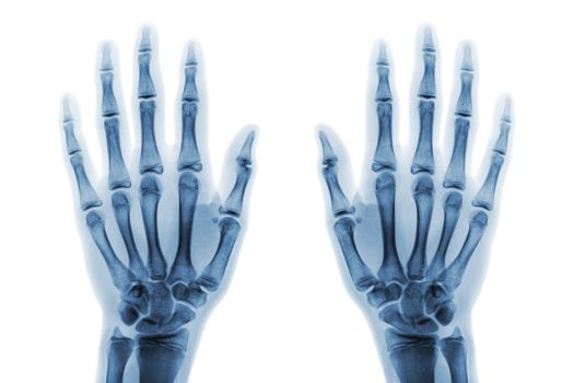
X-ray normal human hands on white background . Oblique view
Stock PhotoUsername
stockdevilResolution
5184x3456pxX-ray normal human hands on white background . Oblique view
X-ray normal human hands on white background . Oblique view
Stock PhotoUsername
stockdevilResolution
5184x3456pxX-ray normal human hands on white background . Oblique view
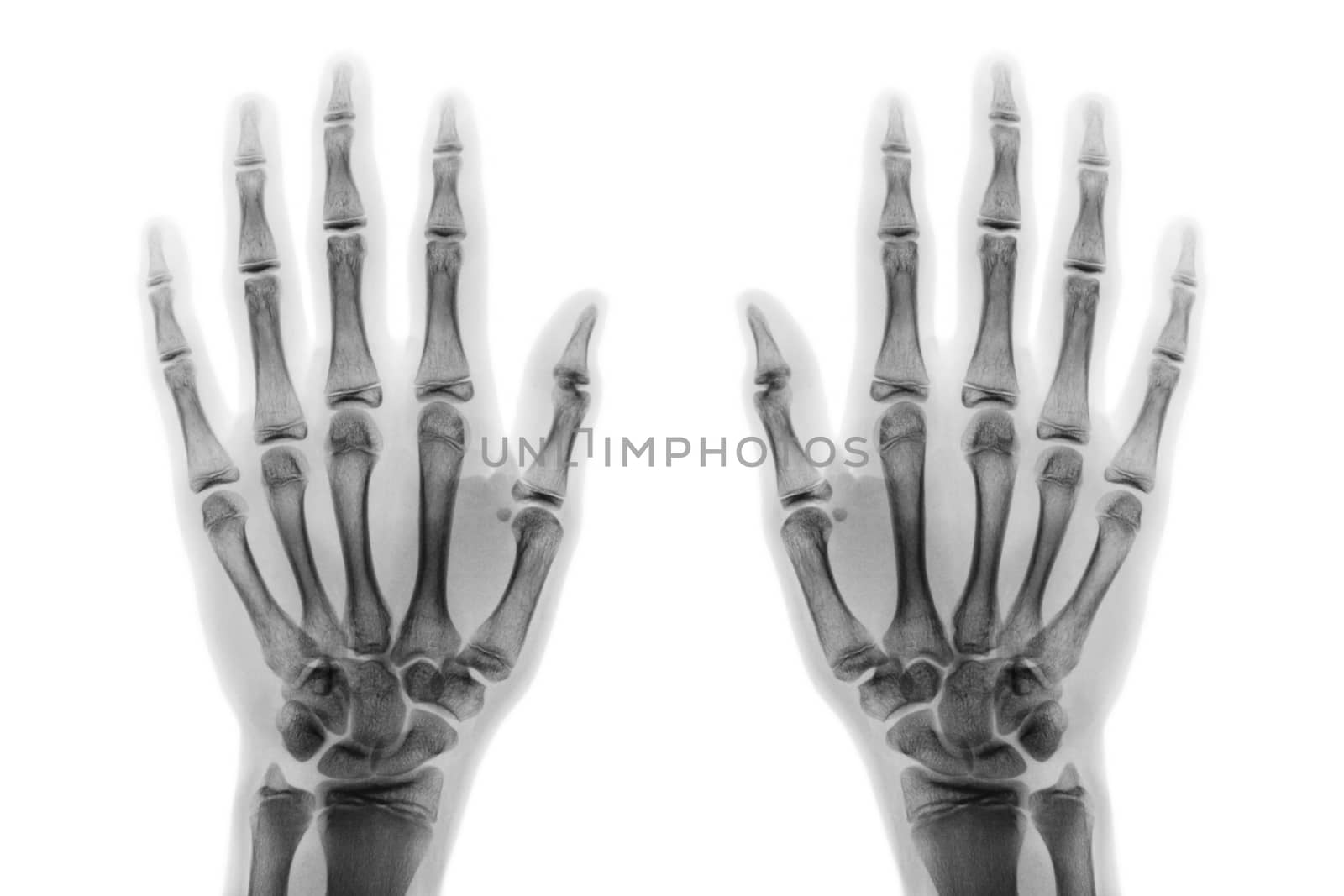
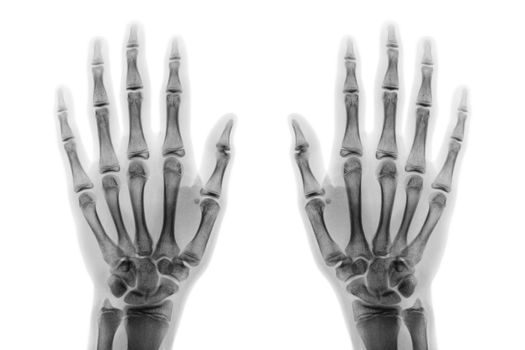
Film x-ray both hand AP show normal human hands on white background ( isolated )
Stock PhotoUsername
stockdevilResolution
5184x3456pxFilm x-ray both hand AP show normal human hands on white background ( isolated )
Film x-ray both hand AP show normal human hands on white background ( isolated )
Stock PhotoUsername
stockdevilResolution
5184x3456pxFilm x-ray both hand AP show normal human hands on white background ( isolated )

Lobar Pneumonia
Stock PhotoUsername
stockdevilResolution
5184x3456pxLobar Pneumonia
Lobar Pneumonia
Stock PhotoUsername
stockdevilResolution
5184x3456pxLobar Pneumonia

Lobar Pneumonia
Stock PhotoUsername
stockdevilResolution
5184x3456pxLobar Pneumonia

Lobar Pneumonia
Stock PhotoUsername
stockdevilResolution
5184x3456pxLobar Pneumonia
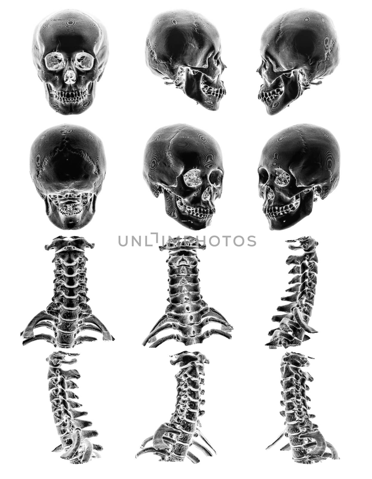
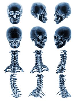
CT scan ( Computed tomography ) with 3D graphic show normal human skull and cervical spine
Stock PhotoUsername
stockdevilResolution
3406x4378pxCT scan ( Computed tomography ) with 3D graphic show normal human skull and cervical spine

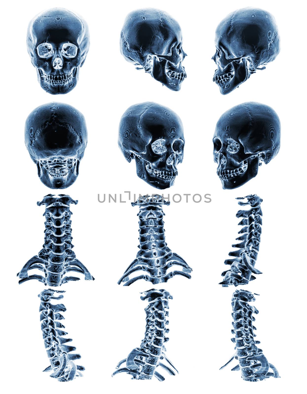
CT scan ( Computed tomography ) with 3D graphic show normal human skull and cervical spine
Stock PhotoUsername
stockdevilResolution
3406x4378pxCT scan ( Computed tomography ) with 3D graphic show normal human skull and cervical spine
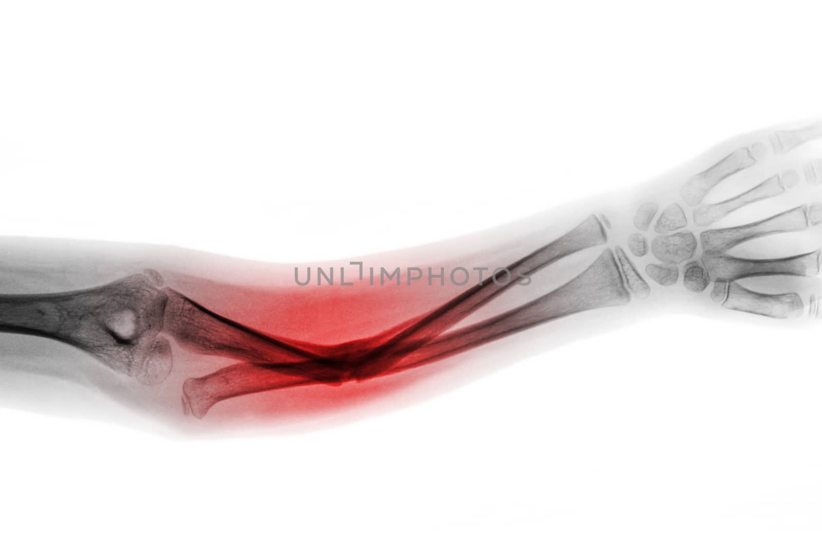
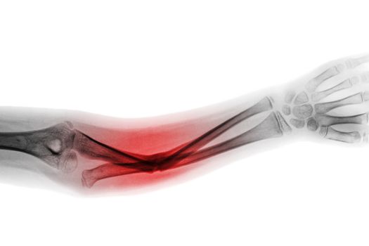
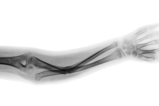
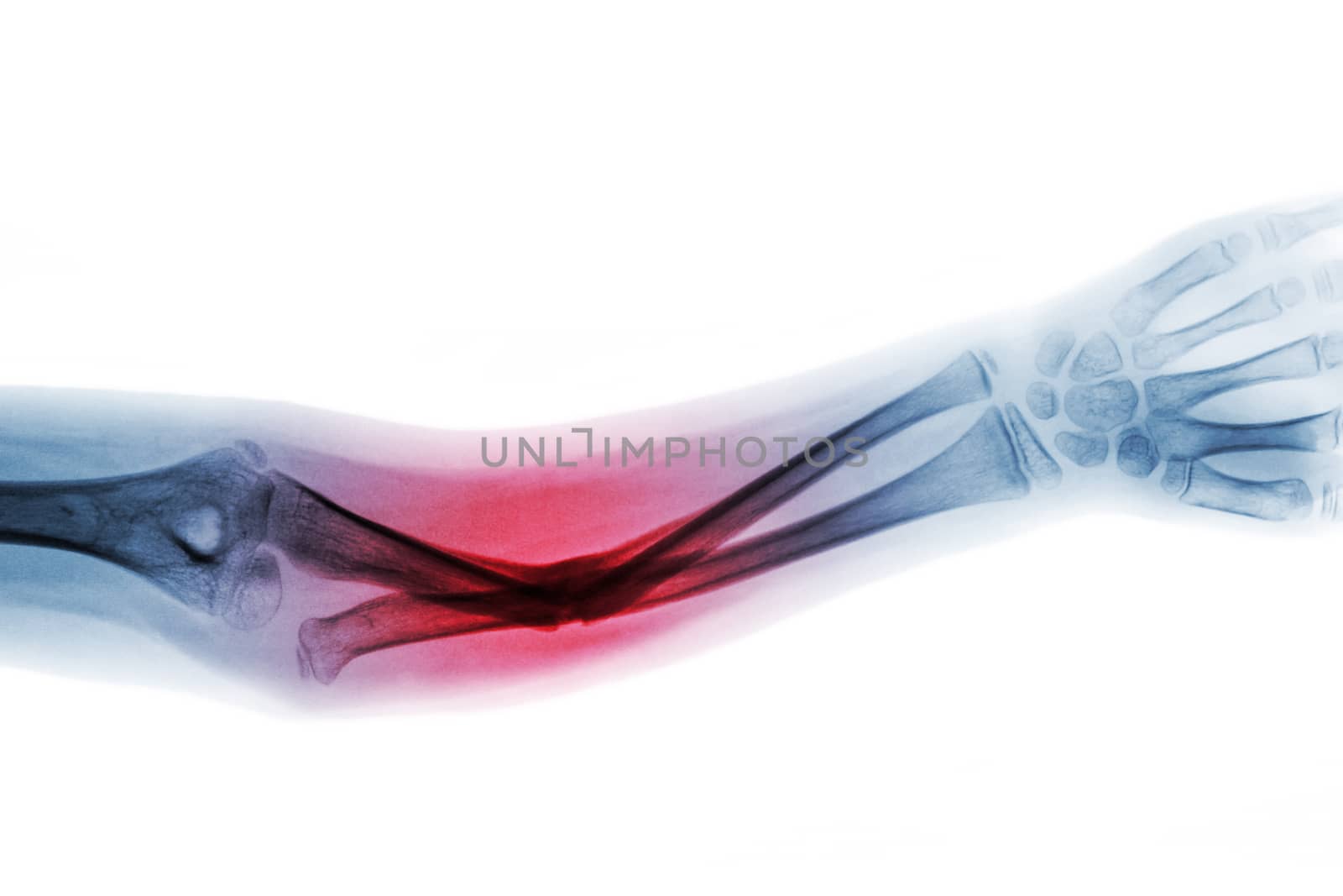
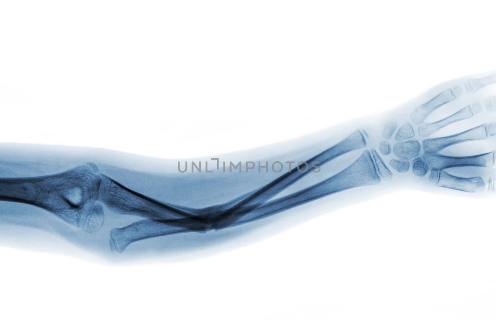
Film x-ray forearm AP show fracture shaft of ulnar bone
Stock PhotoUsername
stockdevilResolution
5184x3456pxFilm x-ray forearm AP show fracture shaft of ulnar bone

Film x-ray forearm AP show fracture shaft of ulnar bone
Stock PhotoUsername
stockdevilResolution
5184x3456pxFilm x-ray forearm AP show fracture shaft of ulnar bone
Film x-ray forearm AP show fracture shaft of ulnar bone
Stock PhotoUsername
stockdevilResolution
5184x3456pxFilm x-ray forearm AP show fracture shaft of ulnar bone

Film x-ray forearm AP show fracture shaft of ulnar bone
Stock PhotoUsername
stockdevilResolution
5184x3456pxFilm x-ray forearm AP show fracture shaft of ulnar bone

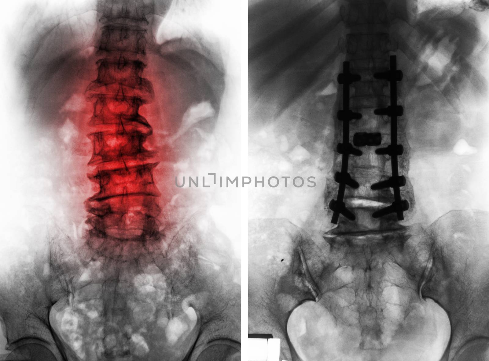
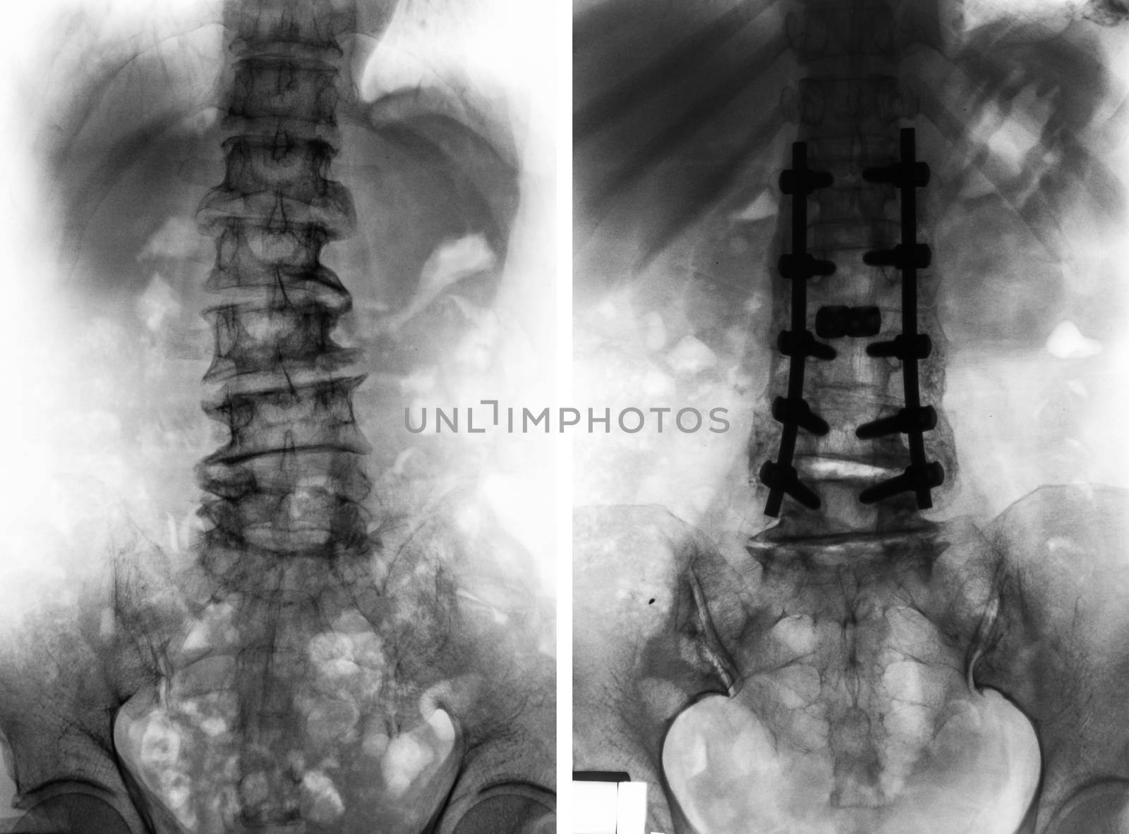
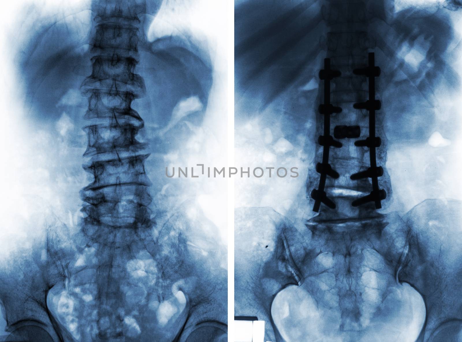
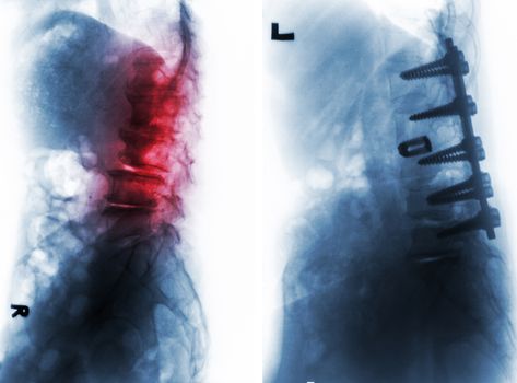
Spondylosis . Before and After surgery
Stock PhotoUsername
stockdevilResolution
7012x5184pxSpondylosis . Before and After surgery
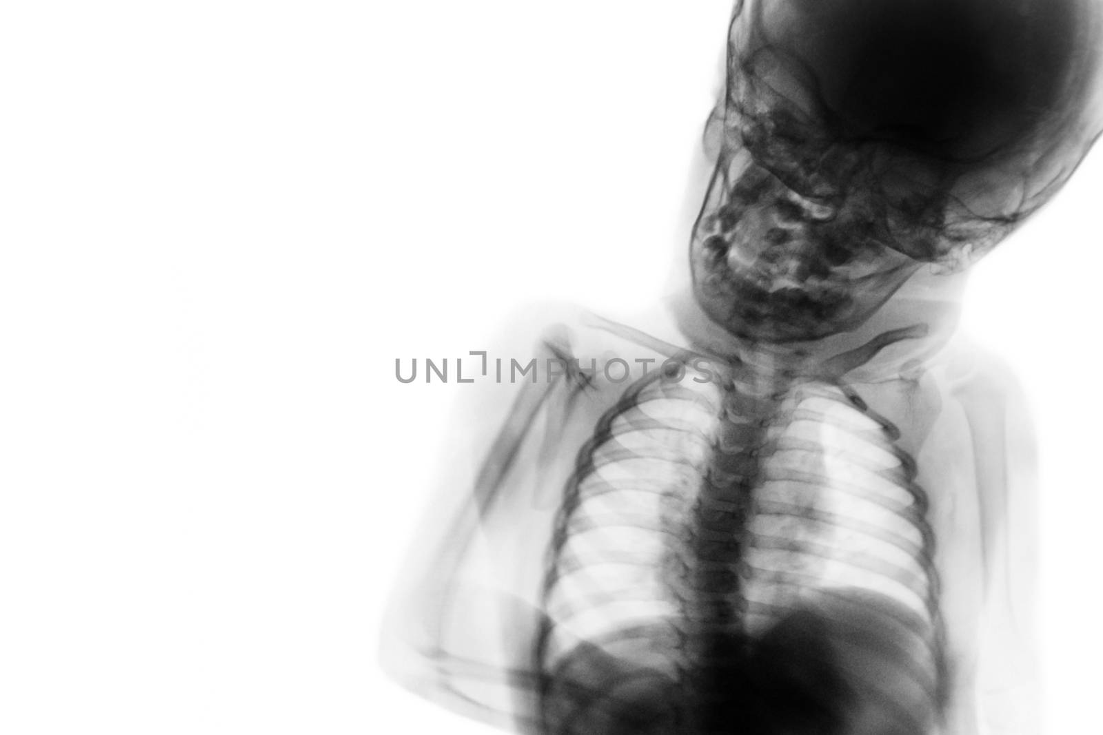
X-ray upper half body of child and blank area at left side
Stock PhotoUsername
stockdevilResolution
5184x3456pxX-ray upper half body of child and blank area at left side
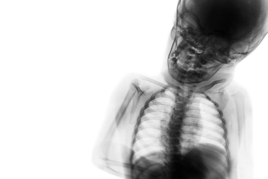
X-ray upper half body of child and blank area at left side
Stock PhotoUsername
stockdevilResolution
5184x3456pxX-ray upper half body of child and blank area at left side
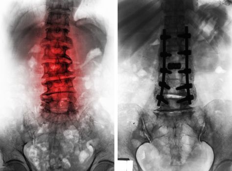
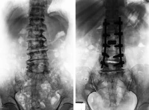
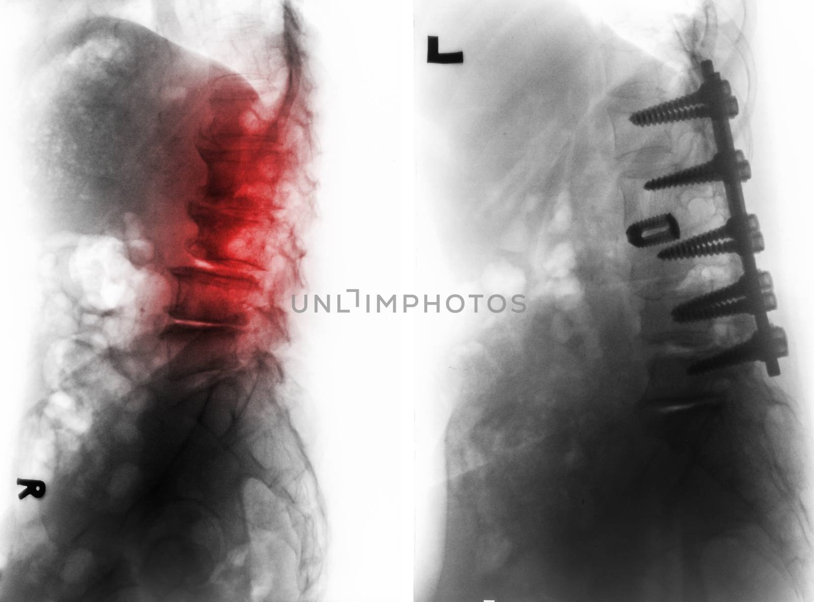
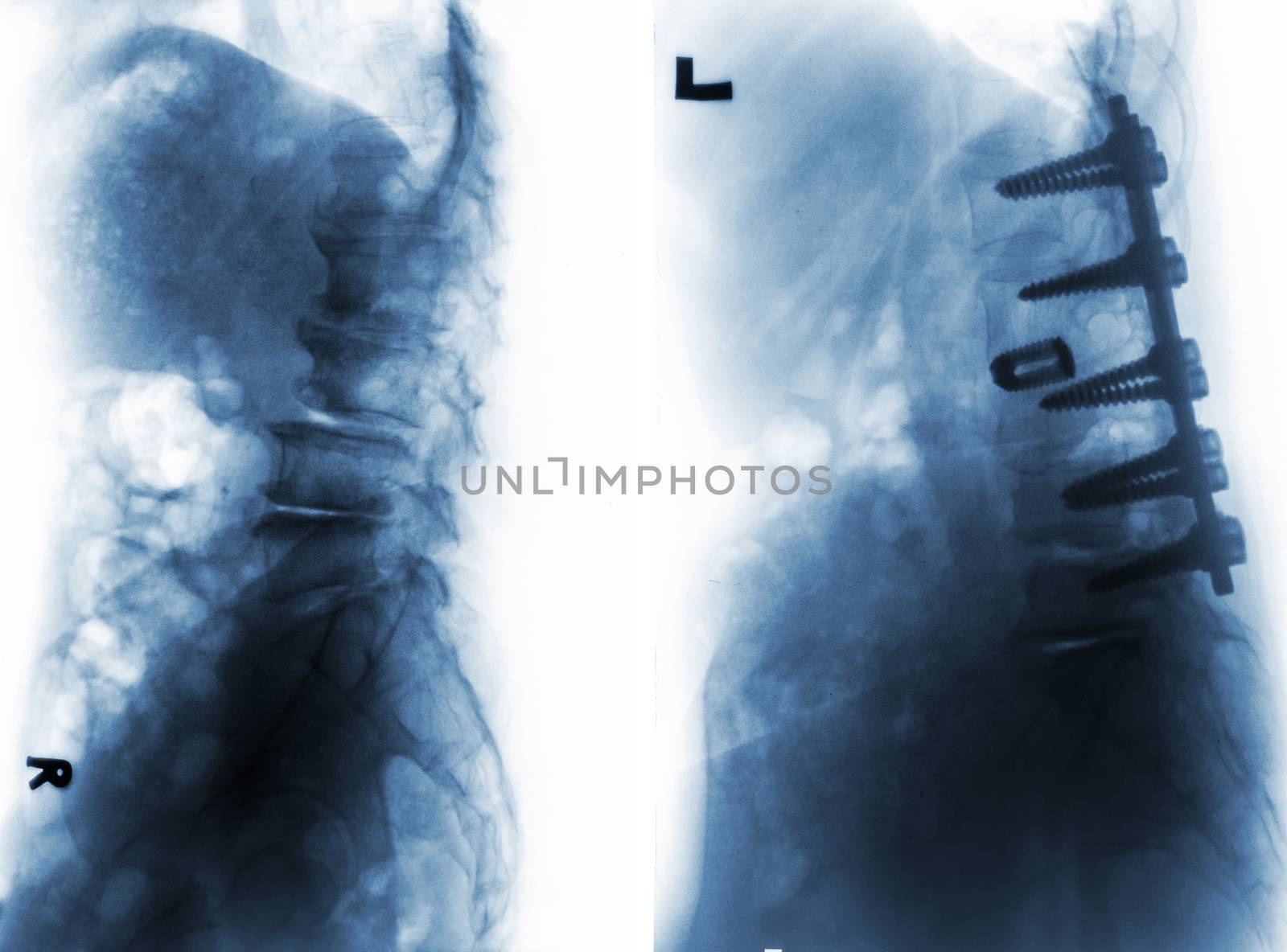
Spondylosis . Before and After surgery .
Stock PhotoUsername
stockdevilResolution
7012x5184pxSpondylosis . Before and After surgery .
Spondylosis . Before and After surgery .
Stock PhotoUsername
stockdevilResolution
7012x5184pxSpondylosis . Before and After surgery .
Spondylosis . Before and After surgery .
Stock PhotoUsername
stockdevilResolution
7012x5184pxSpondylosis . Before and After surgery .

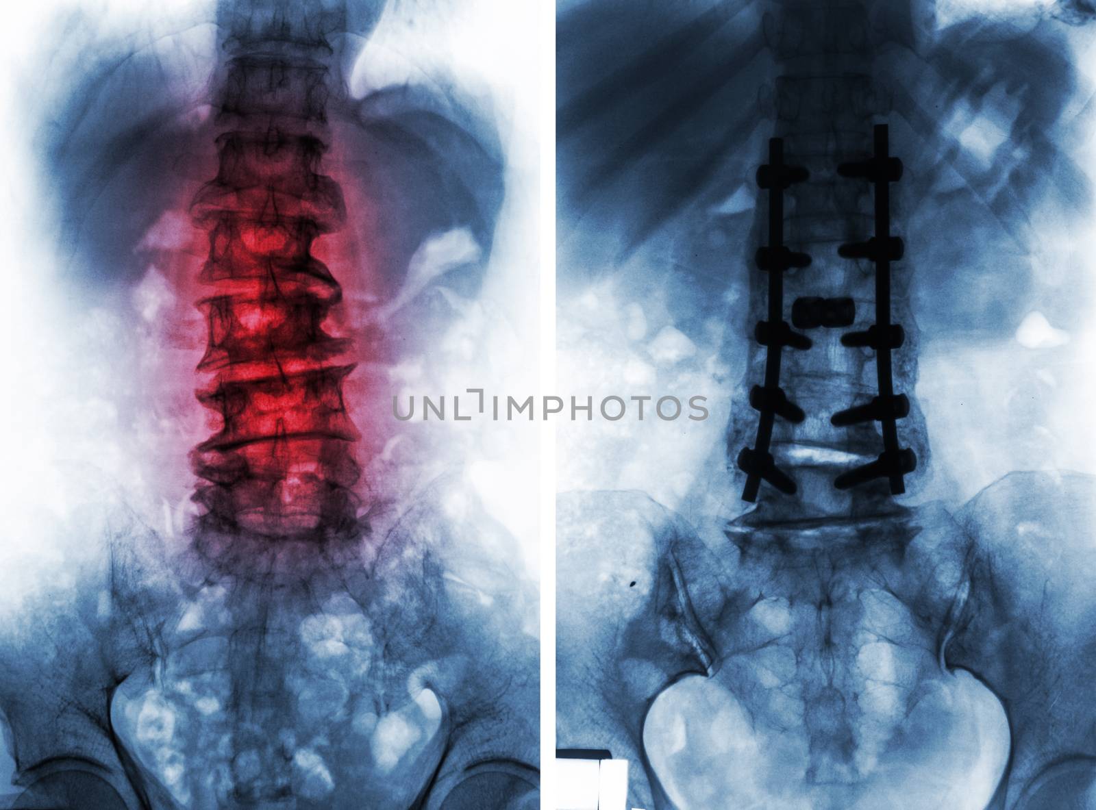
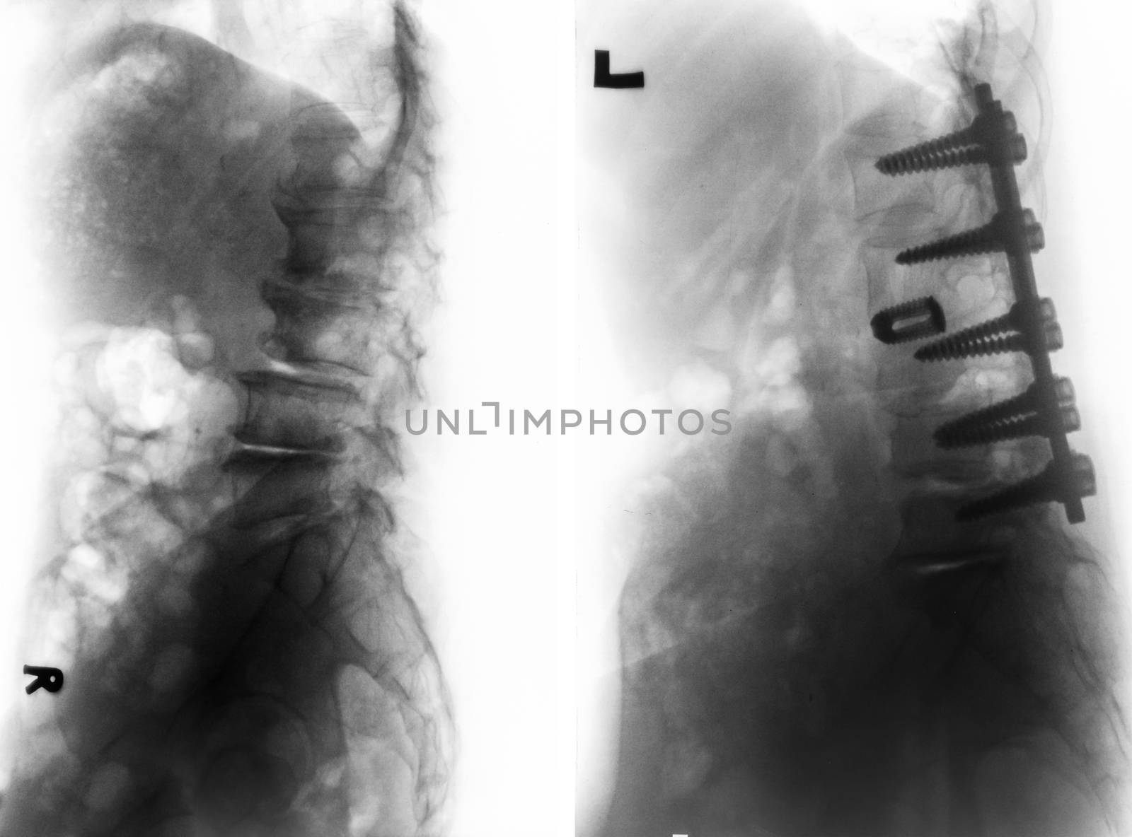
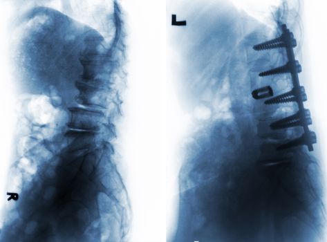
Spondylosis . Before and After surgery .
Stock PhotoUsername
stockdevilResolution
6380x4723pxSpondylosis . Before and After surgery .

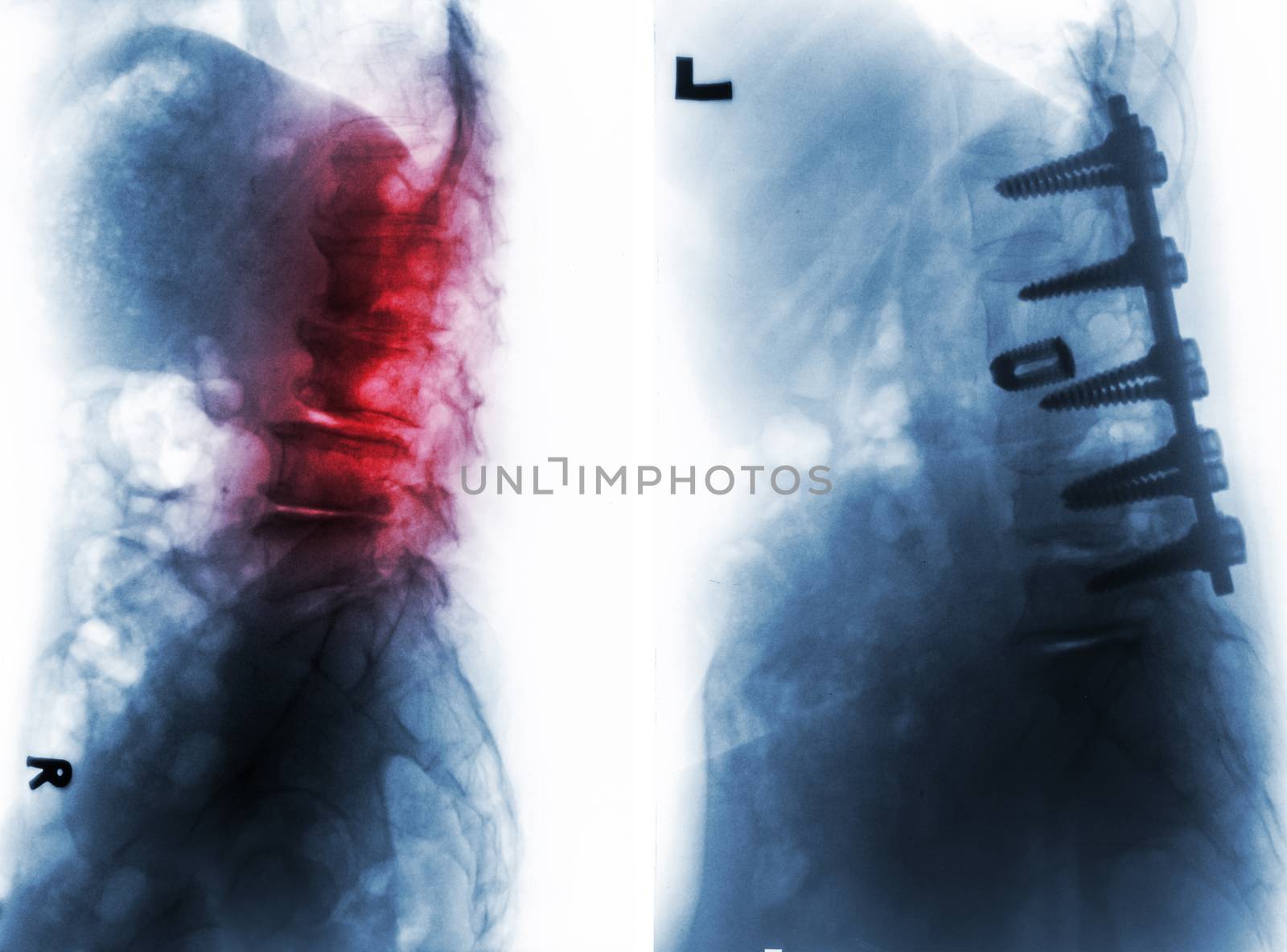
Spondylosis . Before and After surgery .
Stock PhotoUsername
stockdevilResolution
6380x4723pxSpondylosis . Before and After surgery .

Spondylosis . Before and After surgery .
Stock PhotoUsername
stockdevilResolution
6380x4723pxSpondylosis . Before and After surgery .
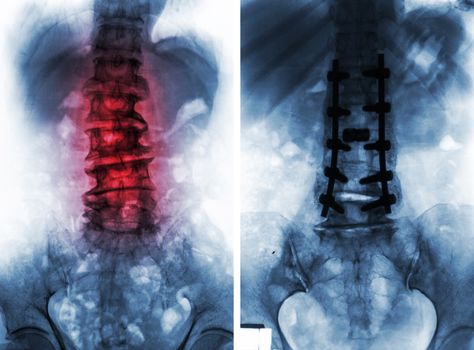
Spondylosis . Before and After surgery .
Stock PhotoUsername
stockdevilResolution
6380x4723pxSpondylosis . Before and After surgery .

Turkey and Greece flag on cracked ground . Confliction and crisis concept .
Stock PhotoUsername
stockdevilResolution
5184x3456pxTurkey and Greece flag on cracked ground . Confliction and crisis concept .


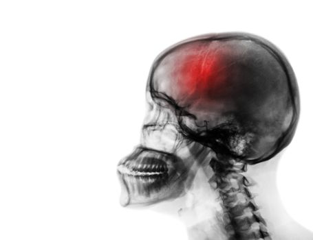
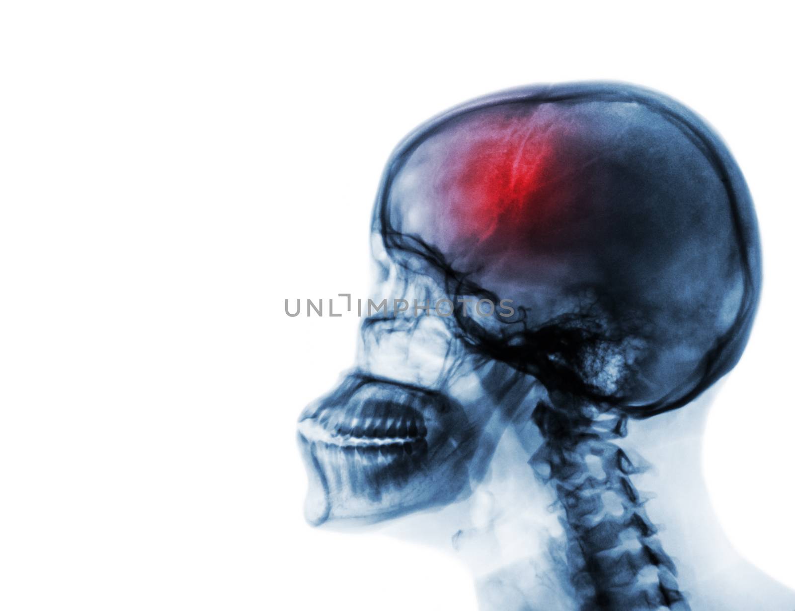
Stroke . Cerebrovascular accident . Film x-ray of human skull and cervical spine .
Stock PhotoUsername
stockdevilResolution
5200x4000pxStroke . Cerebrovascular accident . Film x-ray of human skull and cervical spine .
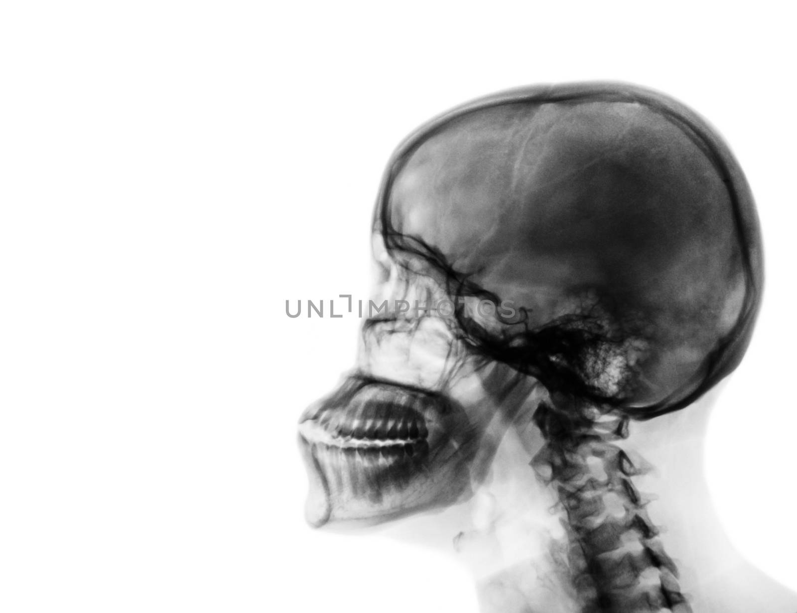
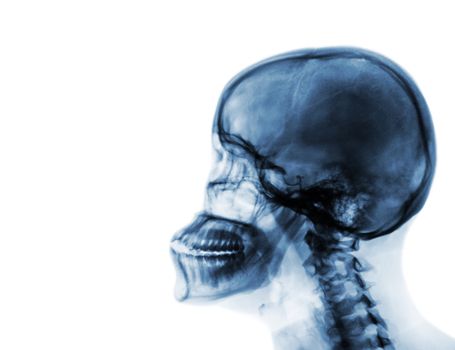
X-ray normal skull and cervical spine . Lateral view .
Stock PhotoUsername
stockdevilResolution
5200x4000pxX-ray normal skull and cervical spine . Lateral view .
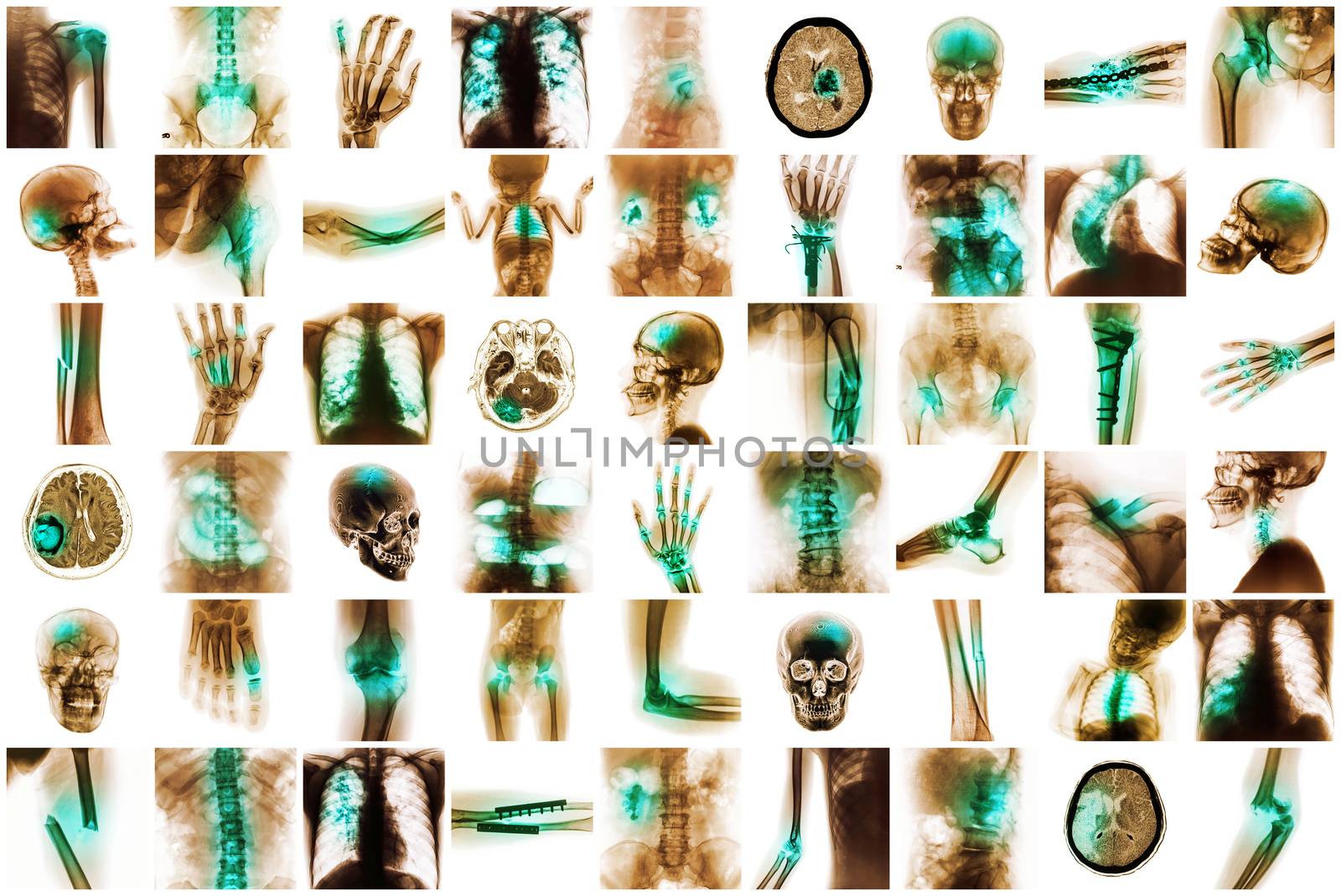
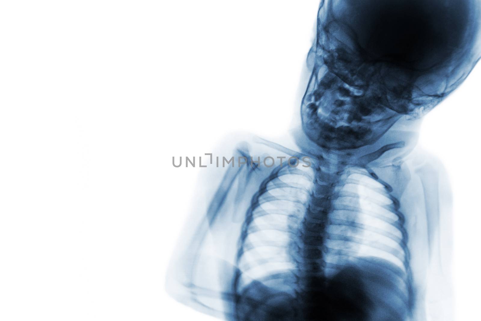
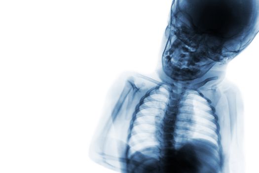
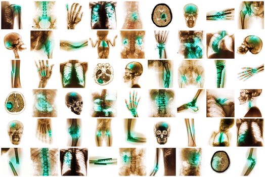
X-ray multiple disease of child and adult
Stock PhotoUsername
stockdevilResolution
6150x4110pxX-ray multiple disease of child and adult
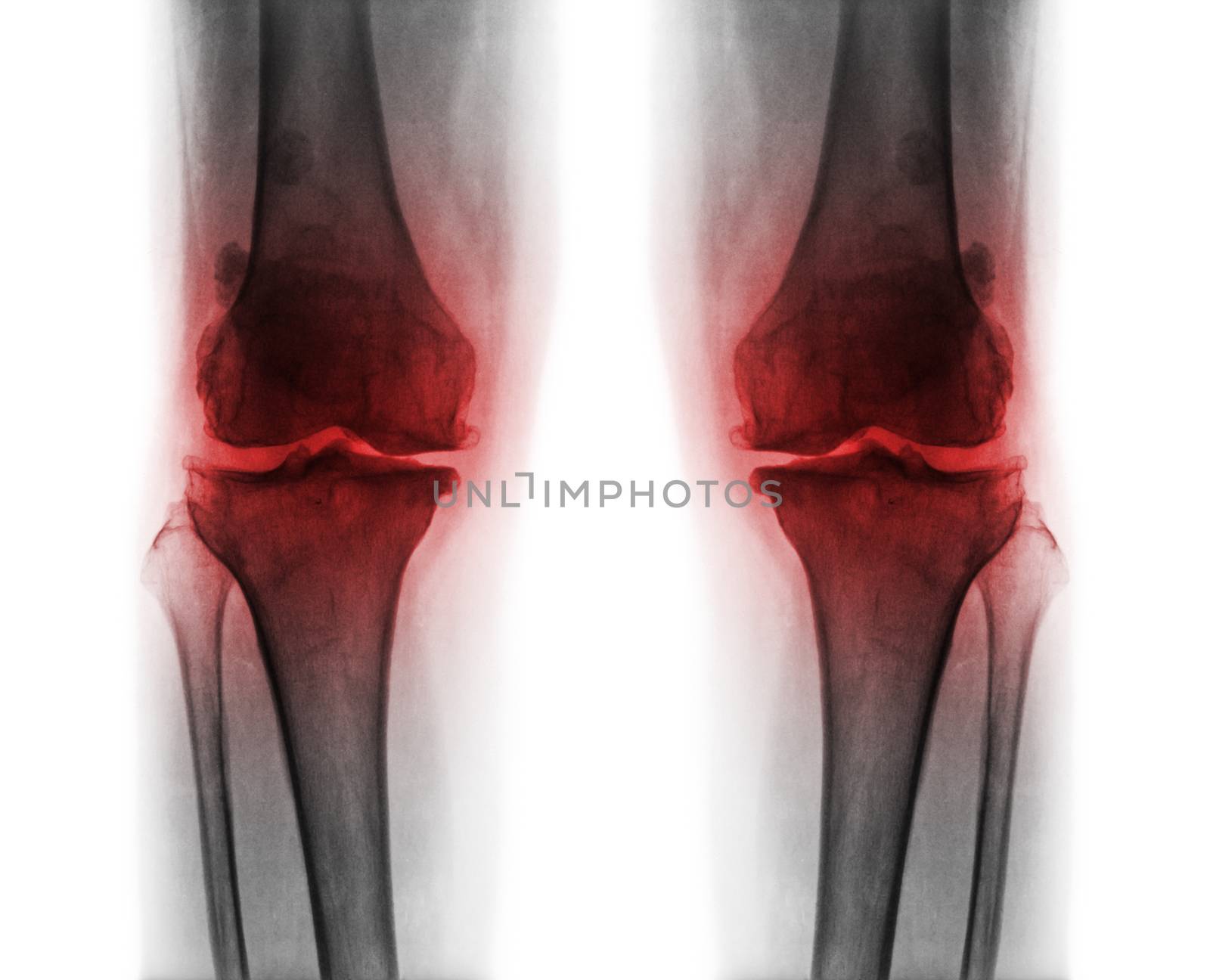
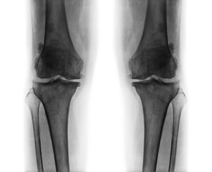
Osteoarthritis both knee .
Stock PhotoUsername
stockdevilResolution
4027x3263pxOsteoarthritis both knee .
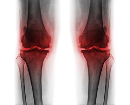
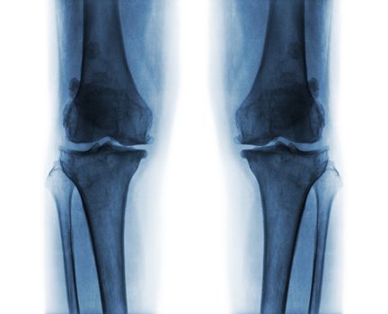
Osteoarthritis both knee .
Stock PhotoUsername
stockdevilResolution
4027x3263pxOsteoarthritis both knee .

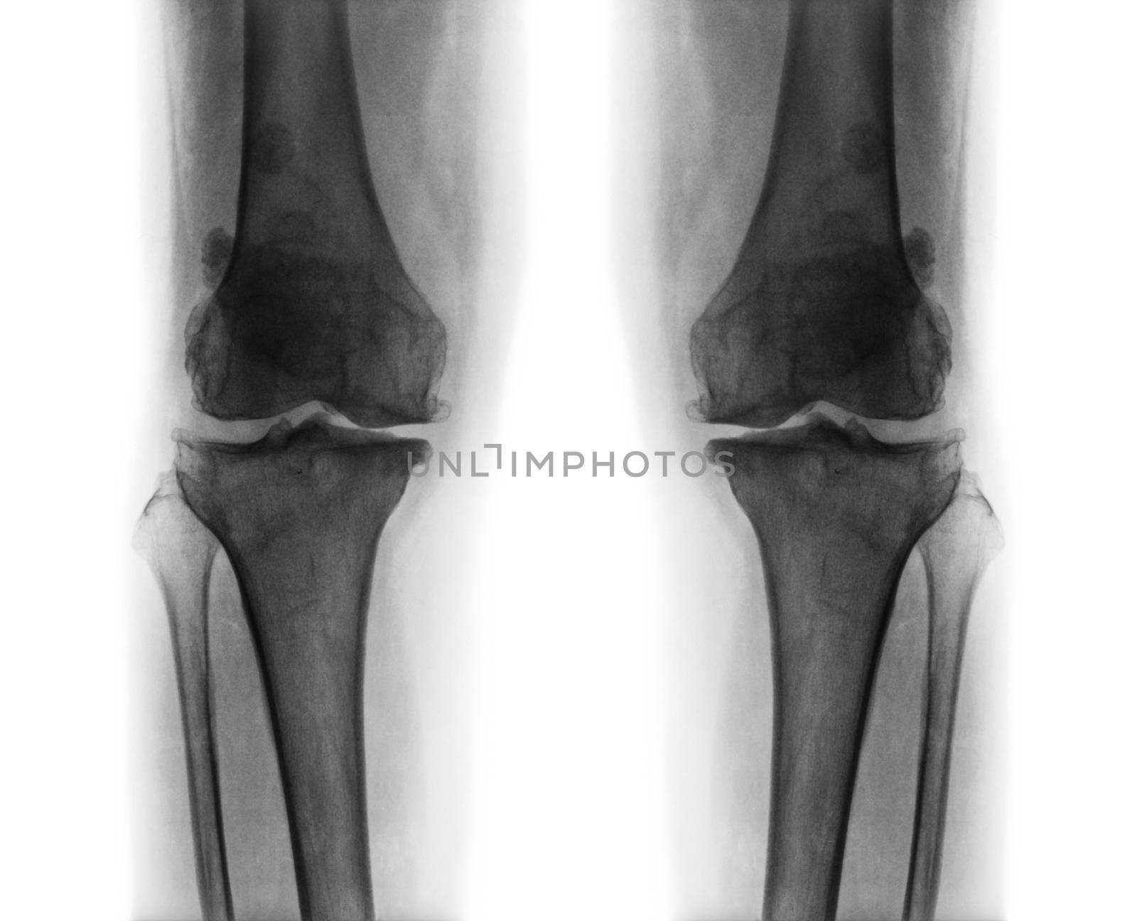
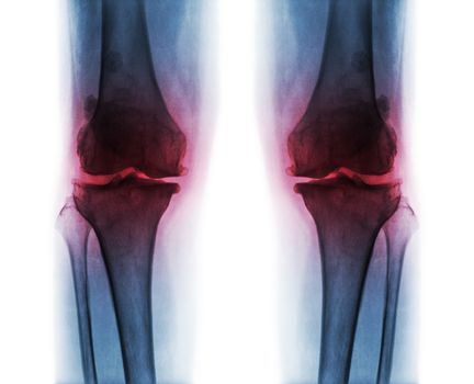
Osteoarthritis both knee .
Stock PhotoUsername
stockdevilResolution
4027x3263pxOsteoarthritis both knee .
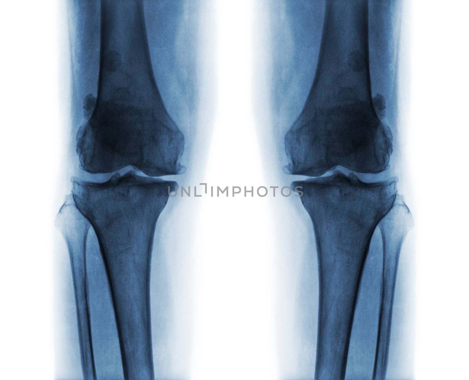
Osteoarthritis both knee .
Stock PhotoUsername
stockdevilResolution
4027x3263pxOsteoarthritis both knee .
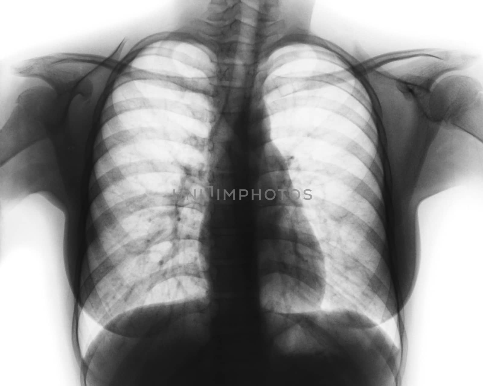
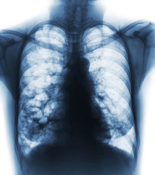
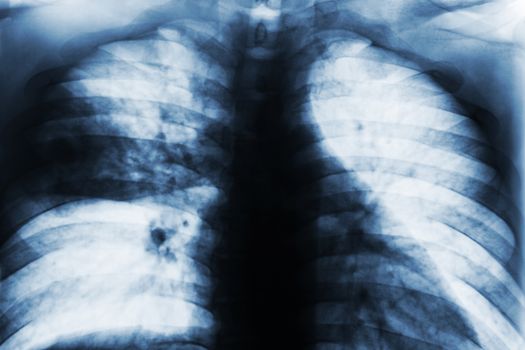
Film chest x-ray of normal woman chest
Stock PhotoUsername
stockdevilResolution
3239x2591pxFilm chest x-ray of normal woman chest
